Medical expert of the article

New publications
Rhinosinusitis in children
Last reviewed: 29.06.2025

All iLive content is medically reviewed or fact checked to ensure as much factual accuracy as possible.
We have strict sourcing guidelines and only link to reputable media sites, academic research institutions and, whenever possible, medically peer reviewed studies. Note that the numbers in parentheses ([1], [2], etc.) are clickable links to these studies.
If you feel that any of our content is inaccurate, out-of-date, or otherwise questionable, please select it and press Ctrl + Enter.
Sinusitis, or the more modern medical definition, rhinosinusitis in children, is a disease of the perinasal sinuses (sinuses) and the natural drainage pathways of the nasal cavity associated with them, with inflammation and swelling of the mucous membrane lining them. The combined term "rhinosinusitis" was coined in 1997 by the Rhinology Working Group and the Committee on the Paranasal Sinuses because sinusitis is invariably accompanied by rhinitis. [1]
Epidemiology
Rhinosinusitis is a common condition that affects more than 14% of adults and children. [2], [3] According to clinical studies, 5-12% of pediatric viral upper respiratory tract infections between the ages of 1 and 5 years progress to acute or chronic rhinosinusitis/sinusitis. Meanwhile, acute bacterial inflammation accounts for approximately 7.5% of cases and occurs most commonly in children aged 4-7 years.
In young children, the maxillary and laryngeal perirhinal cavities are most commonly affected, while in older children and adolescents, either sinus may be inflamed.
The prevalence of deviated nasal septum in chronic rhinosinusitis is estimated at 38-44%. More than 75% of children with rhinosinusitis have a family history of allergy, and more than 50% of cases of paranasal cavity inflammation are associated with asthma.
Causes of the rhinosinusitis in children
Sinusitis in a child can occur:
- Viruses are the most common cause of acute rhinosinusitis, [4] due to frequent colds - in acute lesions of the upper respiratory tract with viral infection (influenza, rhino and adenoviruses), which manifest as rhinopharyngitis;
- In hypertrophy of adenoid vegetations (pharyngeal tonsils) and their inflammation - adenoiditis in children - with spread of bacterial infection (Streptococcus pneumoniae, Streptococcus pyogenes, Pseudomonas aeruginosa, Haemophilus influenzae, Moraxella catarrhalis) into the paranasal sinuses; [5], [6], [7]
- Due to lymphogenic spread of infection from decayed teeth or inflammation of the periosteum of the upper jaw;
- Parasites as a cause of sinusitis are extremely rare and are often found in people with immune disorders; [8]
- As a complication of allergic rhinitis in children;
- In the presence of nasal polyps in a child.
Chronic sinusitis /rhinosinusitis and purulent rhinosinusitis of the paranasal sinuses - maxillary (maxillary), frontal (frontal), cuneiform (sphenoid) or lattice (ethmoid) - may be a consequence of genetically determined cystic fibrosis - hereditary cystic fibrosis in children, as well as kartagener's syndrome, a dysfunction of the cilia (primary ciliary dyskinesia) of the nasopharyngeal mesenteric epithelium.
Also read - what causes acute sinusitis in children?
Risk factors
Risk factors for sinusitis/rhinosinusitis in childhood include:
- Weak immune system or taking medications that weaken the immune system; [9]
- Nasal trauma and nasal foreign bodies;
- Seasonal allergies in children, and respiratory allergies;
- Allergic asthma in children;
- Presence of such anomalies or variants of anatomical structures as deviated nasal septum, unilateral or bilateral bullous (pneumatized) middle nasal concha (concha nasalis media) - conchobullosis, paradoxically curved middle nasal concha (capable of blocking the middle nasal passage), as well as hypertrophy of the hook-like processus uncinatus (processus uncinatus) of the middle nasal concha that directs air into the paranasal sinuses; [10]
- GERD - gastroesophageal reflux disease in children, which may be accompanied by an otolaryngologic (extraesophageal) syndrome in the form of inflammation of the perinasal cavities; [11]
- Periodontitis / a serious dental disease affecting the upper teeth, causing 5-10% of acute rhinosinusitis; [12]
- Swimming, diving, high-altitude rock climbing and diabetes predispose to rhinosinusitis. [13]
Pathogenesis
Specialists note the multifactorial mechanism of rhinosinusitis of any localization and the special importance of the correlation between mucociliary clearance and the state of the ostiomeatal complex (ostiomeatal complex) - a common channel for drainage and ventilation of paranasal sinuses (paranasal sinuses).
These air-filled cavities, lined with ciliated columnar epithelium, communicate through small tubular openings (sinus ostia) that exit into different parts of the nasal cavity. Exocrinocytes (bocaloid cells) of the sinus epithelium produce mucus (mucin), which is transported through the ostiomeatal complex into the nasal cavity due to synchronous oscillatory movement of cilia, called mucociliary clearance. [14]
In inflammatory processes in paranasal sinuses (which are caused by the reaction of immunocompetent cells - neutrophils) due to edema and expansion of the volume of interstitial (extracellular) matrix there is not only hyperplasia of epithelial exocrinocytes, but also narrowing of the sinus mouths and ostiomeatal complex, which causes stagnation of mucous secretion and lack of ventilation of the affected sinus.
The pathogenesis is discussed in more detail in the publications:
Symptoms of the rhinosinusitis in children
In rhinosinusitis, the first signs are a runny nose and nasal congestion (with difficulty or inability to fully breathe through the nose).
Acute rhinosinusitis involves the sudden onset of two or more of the following symptoms: nasal discharge, nasal congestion or stuffy nose, facial pain/pressure, or anosmia/hyposmia. [16], [17] May be accompanied by fever, malaise, irritability, headache, toothache, or cough. If symptoms persist for 4-12 weeks, it is subacute rhinosinusitis. When they persist for more than 12 weeks, it is called "chronic rhinosinusitis." [18] The latter usually results from untreated/incorrectly treated/refractory acute rhinosinusitis. Recurrent rhinosinusitis is 4 or more episodes of acute sinus infection in one year, each lasting about a week. By etiology, rhinosinusitis can be viral, bacterial, fungal, parasitic, or mixed.
Nasal discharge in the initial stage of catarrhal inflammation has a serous character (they are transparent and watery). But then the discharge becomes thick, muco-purulent - yellow or greenish in color. See - purulent rhinitis
Common symptoms of sinus infection include: decreased sense of smell, aching or throbbing facial pain and pressure/spreading sensation in the face, headache, ear and jaw pain, postnasal congestion of mucus (in the throat), sore throat and cough, and bad breath.
Often noted chills and fever in rhinosinusitis in a child, especially younger children.
In cases of acute rhinosinusitis with localization in the lattice sinus (ethmoiditis) pain of a pressing nature occurs deep in the space between the eyebrows, in the bridge of the nose and the inner corner of the eye, there is an increased lacrimation, redness of the conjunctiva of the eyes and swelling of the eyelids.
Also read:
Forms
The sinuses - air-bearing cavities within the skull that connect to the nasal cavity - are located in the three bones of the cerebral skull (neurocranium): frontal (os frontale), lattice (os ethmoidale) and cuneiform (os sphenoidale); the maxillary sinus is located in the maxilla bone of the facial part of the skull (viscerocranium). The maxillary and lattice sinuses are formed during intrauterine development; the cuneiform sinuses appear in the fifth month of postnatal life, and development of the frontal sinus begins at the age of two years.
According to the localization of the inflammatory process, such types or kinds of rhinosinusitis/sinusitis are distinguished as:
- Maxillary sinusitis/rhinosinusitis (inflammation of the maxillary or maxillary sinus);
- Frontal rhinosinusitis/sinusitis (inflammation of the frontal, i.e. Frontal sinus);
- Sphenoidal rhinosinusitis/sinusitis (inflammation of the cuneiform or sphenoid sinus);
- Ethmoidal or lattice sinusitis or rhinosinusitis.
If the symptoms do not appear for more than four weeks, it can be defined as acute rhinosinusitis in children or acute catarrhal rhinosinusitis in a child. And if there is pus in the paranasal cavity and its presence in the nasal discharge - acute purulent rhinosinusitis in a child, and, as a rule, it is bacterial rhinosinusitis.
When sinus inflammation was preceded by acute respiratory viral infections, the ENT doctor can establish post-viral rhinosinusitis in a child. Since viral infection is associated with increased bacterial growth, the development of secondary bacterial inflammation cannot be ruled out.
Recurrent or recurrent rhinosinusitis may develop with frequent respiratory illnesses.
Read more in the publications:
- Acute sinusitis in children
- Acute maxillary sinusitis (maxillary sinusitis)
- Acute frontitis
- Acute ethmoidosphenoiditis.
- Acute inflammation of the labyrinth (acute rhinoethmoiditis)
When symptoms last longer, chronic rhinosinusitis in a child is defined:
If polyps are found in the perinasal cavity that narrow their drainage vessels, chronic polyposis rhinosinusitis in children is diagnosed.
It is clear that the presence of seasonal allergies or allergic asthma gives the otolaryngologist and allergist every reason to define inflammation of any paranasal sinus as allergic rhinosinusitis in children. And simultaneous inflammation of both paired sinuses will be diagnosed as bilateral rhinosinusitis in a child.
Complications and consequences
Sinusitis/rhinosinusitis in children can become complicated:
- Formation of mucocele of the sinuses (most often in the frontal and lattice sinuses);
- Inflammation of the eustachian (auditory) tube and the development of chronic otitis media;
- Empyema (pus buildup) of the posterior cells of the lattice sinus;
- Formation of an oroantral fistula - a pathologic fistula between the oral cavity and the maxillary sinus;
- Meningitis or arachnoiditis, an inflammation of the soft and webbed membranes of the brain;
- With a brain abscess;
- Isolated paralysis of the oculomotor nerves, retrobulbar neuritis, inflammation of the lacrimal ducts, inflammation of the ocular vasculature (chorioiditis) with accession of inflammation of the retina (chorioretinitis) and other rhinogenic ophthalmologic complications;
- Osteomyelitis of the facial bone structures of the skull.
Diagnostics of the rhinosinusitis in children
Crucial for determining the tactics of therapy of rhinosinusitis is the correct diagnosis, the basis of which are: [19]
- History, physical findings and clinical manifestations;
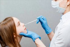
- Instrumental diagnostics, such as an anterior rhinoscopy, endoscopy (examination) of the nasal cavity, ultrasonography (ultrasound), [20] x-rays of the nasal cavity and paranasal sinuses or CT scans of the appendicular cavities; [21]
- Tests (general blood test and IgE antigen, nasal mucus test ). [22]
Read more:
Since the symptoms of sinusitis/rhinosinusitis are similar to the clinical manifestations of other pathological conditions, differential diagnosis is very important - with adenoiditis, cysts and other neoplasms of the nasal cavity and paranasal sinuses.
Who to contact?
Treatment of the rhinosinusitis in children
Therapy of rhinosinusitis/sinusitis in pediatric practice does not differ much from the treatment of this disease in adults.
Acute rhinosinusitis usually resolves on its own and recovers with symptomatic treatment and minimal intervention. Steam inhalation, adequate hydration, injection of topical anti-inflammatory agents, application of warm face masks and saline nasal drops are useful. Elevation of the head during sleep brings relief. Nasal decongestants reduce mucus production and can be safely used for 5-7 days. Prolonged use beyond this period may lead to recurrent vasodilation and worsening of nasal congestion. [23] However, a study by McCormick et al. Found no benefit of a topical antitussive agent with an oral antihistamine in children with acute rhinosinusitis. [24] Nasal saline irrigations, nasal steroids, and topical cromolyn were found to be helpful. Saline irrigations promote mechanical clearance of secretion, minimize bacterial and allergen load, and improve mucociliary function. [25] Nasal steroid drops or cromolyn drops or sprays improve symptoms in children with concomitant nasal allergies. A short course of systemic steroids is used before surgery to minimize intraoperative blood loss in children with nasal polyps. [26] Antihistamines are helpful for people with concomitant nasal allergies. But they tend to thicken secretions and further aggravate rhinitis and orifice obstruction. It has been observed that mucolytics have different effects. Adequate randomized and controlled trials to evaluate their efficacy in such patients have not been conducted. [27], [28] The use of antibiotics is usually not justified. A "wait-and-see" policy of 7-10 days is fruitful and cost-effective. About 90% recover without antibiotics within a week. [29] Antibiotics are prescribed for children with severe acute sinusitis, toxic manifestations, suspected complications, or persistence of symptoms. [30] The choice of antibiotics should be based on the results of local sensitivity studies, safety profile, and age of the child. Amoxicillin, co-amoxiclav, oral cephalosporins and antibiotics of the macrolide group are usually preferred. A 2-week course is usually required. [31]
Details:
Which medicines are used, read in the articles:
- Antibiotics for sinusitis
- Treatment of frontitis with antibiotics
- Drops for maxillary sinusitis
- Sprays for maxillary sinusitis
- Nasal spray for children
- Nasal rinsing for maxillary sinusitis
- Nasal rinse for a baby
- Nasal rinses
In allergic rhinosinusitis in children, systemic antihistamines and intranasally - sprays for allergic rhinitis are prescribed.
Physiotherapy treatment is used:
In some cases, primarily when drug therapy is ineffective, surgical treatment is necessary.
In acute and chronic maxillary sinusitis of bacterial origin that does not respond to conservative treatment, the simplest (but largely obsolete) method is to perform an maxillary sinus puncture - a puncture of the maxillary sinus cavity - and lavage (lavage) through a cannula inserted into the maxillary sinus through the lower nasal passage. Several repeated lavages are often required to ensure that the accumulated pus from the infection is completely flushed out.
If the visualized amount of adenoid tissue is found to be sufficient as a reservoir for bacterial infection, surgical intervention in the form of adenoidectomy - adenoid removal in children. Is indicated.
There is also removal of nasal polyps
In a limited anterior ethmoidectomy, infected tissues that block the natural drainage of this cavity are removed from the lattice paranasal sinus.
In cases of anatomical anomalies that need to be corrected, endoscopic surgery of the paranasal sinuses is used. For example, during an unziectomy, the anterior, inferior and superior attachments of the middle nasal hook attachment are separated and removed.
Additionally see. - surgery for chronic maxillary sinusitis
Prevention
Basic medical recommendations for preventing inflammation of the perinasal sinuses are given in the material - prevention of upper respiratory tract infections in children
Forecast
In the case of rhinosinusitis in children, as in the development of this disease in adults, the prognosis is determined by the etiology, localization and success of treatment of inflammation of the paranasal sinuses.
Использованная литература