Medical expert of the article

New publications
Duplex scanning of head and neck vessels
Last reviewed: 29.06.2025

All iLive content is medically reviewed or fact checked to ensure as much factual accuracy as possible.
We have strict sourcing guidelines and only link to reputable media sites, academic research institutions and, whenever possible, medically peer reviewed studies. Note that the numbers in parentheses ([1], [2], etc.) are clickable links to these studies.
If you feel that any of our content is inaccurate, out-of-date, or otherwise questionable, please select it and press Ctrl + Enter.
Among the many instrumental ultrasound diagnostic methods used by cardiologists, neurologists and surgeons, duplex scanning of head and neck vessels is particularly common. This study is based on the properties of ultrasound vibrations reflected from the vascular walls and individual blood cells - red blood cells, which allows you to consider the state of the vessels and assess the quality of blood circulation.
Indications
Separate duplex scanning of brachial and external cranial vessels (these include external carotid, vertebral and subclavian arterial and venous vessels), as well as duplex scanning of cerebral vessels and intracranial vascular network.
Ultrasound type of scanning is available and informative, helps to determine and identify various pathological processes. This diagnostic method is used if the patient complains of regular and pronounced pain in the head, as well as:
- Dizziness, pre-syncope and fainting spells that occur intermittently or frequently;
- Trouble sleeping;
- Intermittent palpitations, heart rhythm failures;
- Blood pressure fluctuations, tendency to high blood pressure;
- Frequent sensation of noise and ringing in the head or ears;
- Frequent nosebleeds;
- The occurrence of a shroud of, "goosebumps" in front of the eyes;
- Deterioration of auditory, visual function;
- Pain in the neck, back of the head for no apparent reason;
- Changes in gait for no reason;
- Sharp deterioration in concentration, memory problems.
In addition, the doctor may prescribe a duplex scan of the vessels of the head and neck for patients:
- After stroke, other acute or chronic forms of circulatory disorders of the brain (including transient ischemic attacks );
- For hypertension, diabetes;
- After myocardial infarctions, in angina pectoris, etc.;
- In diagnosed vascular atherosclerosis;
- When high cholesterol and low-density lipoproteins in laboratory tests;
- At heart defects;
- For cervical osteochondrosis;
- After head trauma (PMT).
Duplex scanning of the vessels of the head and neck is indicated at the preparatory stage before surgery in the brain, as well as in case of suspected tumor process, for dynamic monitoring of the effectiveness of treatment, or to assess the general condition of the body.
Preparation
Preparation for the procedure is not complicated. On the eve of duplex scanning of the head and neck vessels, the patient should refuse smoking, alcohol and psychotropic drugs (about 24 hours before the study).
In addition, it is recommended to refuse taking medications that can affect the cardiovascular system (after consulting with your doctor), do not drink coffee, tea, do not eat 4-5 hours before the procedure.
No other preparations are usually required. In some cases, the doctor may make individual recommendations.
Technique
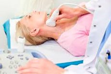
The patient removes outer clothing, undresses to the waist, lies on the couch on the back, or on the right or left side (at the discretion of the doctor). During the procedure it is not recommended to move, talk - only if the doctor asks you to do so.
Before beginning the examination, the doctor measures the patient's blood pressure on the left and right arm.
The ultrasound specialist applies a special gel to the scanning sensor for better contact with the skin, and then applies the device to the lateral cervical surface, the area above the back of the head, above the collarbones, to the temporal area.
If the patient experiences dizziness or other discomfort during the process, he or she should inform the doctor.
What does a duplex scan of the head and neck vessels show?
Thanks to duplex scanning of the head and neck vessels, it is possible to thoroughly examine the condition of the vascular walls associated with the brain and neck region. The doctor is able to assess the features of the main, superficial and deep arterial and venous vessels, determine the degree of their patency, measure the wall thickness.
A duplex scan can detect:
- Narrowing of the vascular lumen;
- Changes in the thickness of the vessel wall, areas of delamination;
- Pathologic lumen dilatations, aneurysms;
- Excessive tortuosity.
The norm for a healthy person is defined by an adequate vascular network with good patency, anatomically correct wall thickness and lumen width. Any pathologic dilatations, delaminations, inclusions and formations should be absent.
Decoding of duplex scanning of head and neck vessels
Deciphering the results of duplex scanning is performed by an ultrasound specialist or attending physician. Standardly assessed is the condition of vessels, patency, the presence of pathologic inclusions in extra intracranial venous and arterial vessels:
- The brachial trunk;
- Subclavian arteries;
- Carotid, vertebral arteries;
- Of the internal jugular veins;
- Anterior, middle cerebral arteries;
- Posterior cerebral arteries;
- The main artery, the anterior and posterior connecting vessels.
To determine the degree of narrowing of the carotid arteries, it is recommended to apply such diagnostic criteria:
- In norm - the ultimate systolic flow velocity through the internal carotid artery should be no more than 125 cm/sec, without visible layering and thickening of the internal vascular layer;
- Constriction 50-69% ultimate systolic velocity - 125-230 cm/sec;
- Constriction exceeds 70%, the systolic velocity limit exceeds 230 cm/sec;
- The narrowing exceeds 90%, a pronounced vascular stenosis is registered, the blood circulation speed is sharply limited.
If there is complete occlusion of the lumen, the blood velocity is not recorded at all.
Additionally, the ratio of the systolic velocity limit in the common and internal carotid artery is assessed. If the internal carotid artery is narrowed, the ratio increases by a factor of 3 or more. This indicator is particularly relevant for patients with heart failure and reduced myocardial (left ventricular) ejection fraction.
With the help of modern technology during duplex scanning of the head and neck, the state of the intima-media complex is determined. This is the inner layer of arteries, where atherosclerotic changes first appear. Thickness indicators, structural features of the intima-media complex are important diagnostic and prognostic values. It is generally accepted that an increase in the intima-media complex thickness in the common carotid artery of more than 0.87 mm (and in the internal carotid artery of more than 0.9 mm) is a marker associated with an increased risk of cardiovascular diseases, including cerebral circulatory disorders and infarcts.
Most often duplex scanning of the vessels of the head and neck reveals signs of atherosclerotic changes - in particular, plaques of different sizes, structure, composition, as well as thrombi. The ultrasound specialist should describe as detailed as possible the picture seen with the localization of the detected changes.