Medical expert of the article

New publications
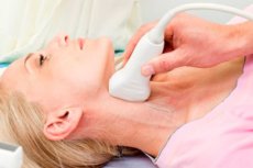
Ultrasound of the brachiocephalic arteries
Last reviewed: 29.06.2025

All iLive content is medically reviewed or fact checked to ensure as much factual accuracy as possible.
We have strict sourcing guidelines and only link to reputable media sites, academic research institutions and, whenever possible, medically peer reviewed studies. Note that the numbers in parentheses ([1], [2], etc.) are clickable links to these studies.
If you feel that any of our content is inaccurate, out-of-date, or otherwise questionable, please select it and press Ctrl + Enter.
If there is a need to assess the state of the vascular network that feeds the brain area, ultrasound of the brachiocephalic arteries is prescribed. This technique allows you to identify problems in the structure of the vascular walls, narrowing of the arteries, causing a deficit of blood supply to the brain tissues. Thanks to ultrasound, it is possible to detect aneurysms, strokes, transient ischemic conditions at the initial stages of development.
What does brachiocephalic artery ultrasound mean?
The condition of the arteries directly affects the quality of the entire body. Vascular problems do not appear suddenly, but progress over time.
Ultrasound examination of the brachiocephalic arteries allows timely detection of even initial pathologic changes.
Ultrasound involves the use of duplex and triplex scans. Both methods are safe and belong to non-invasive diagnostic methods.
Thanks to duplex scanning, it is possible to determine the quality of vascular patency, to find the cause of its violation. The method is based on ultrasound Dopplerography, which evaluates the features of blood flow and its direction.
Duplex scanning provides the doctor with a two-dimensional image of the arterial walls.
Triplex ultrasound of brachiocephalic arteries includes duplex scanning technique and color mode Doppler. Triplex allows you to view the arterial structure and structure, identify the features of blood flow and assess vascular patency in color.
During the ultrasound of the brachiocephalic arteries, any radiation exposure to tissues and organs is excluded, so the study is allowed to undergo, including pregnant women and infants. The ultrasound session itself can be performed at any frequency, depending on the need.
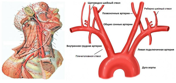
Brachiocephalic arteries include all arterial trunks localized in the cervical segment of the vertebral column. These are the common carotid artery, the left subclavian artery, the brachial trunk. In this case, ultrasound examination involves assessment of the state of extracranial arteries that go to the brain structures and are responsible for supplying them with blood.
Indications for the procedure
This type of examination is prescribed in case of suspected circulatory disorders in the head and neck area.
The main indications are considered to be:
- Severe headaches, not relieved by appropriate medication, migrating;
- Sensation of tinnitus and ringing in the ears, vestibular disorders;
- Visible pulsation of the temporal vessels;
- Changes in gait, wobbling, trouble climbing stairs or getting out of bed;
- Regular dizziness, sometimes to the point of semi-fainting and fainting;
- Blood pressure fluctuations, difference in pressure readings in the right and left arm;
- Sleep disturbance at night against the background of constant daytime sleepiness;
- Upcoming surgical intervention in the cervical spinal column;
- Evaluation of the dynamics of the conducted vascular treatment;
- Monitoring of the postoperative condition.
For preventive purposes, bracheocephalic artery ultrasound may be recommended for patients with hormonal imbalances, systemic pathologies with an increased risk of metabolic complications.
The method is also used to determine the severity of pathological changes in patients with strokes and heart attacks.
Diagnosis usually requires a referral from your primary care physician.
There are no special contraindications to ultrasound of the brachiocephalic arteries. In some cases, dermatologic diseases and skin lesions in the neck area, obesity, mental abnormalities may prevent the diagnostic procedure.
Preparation
How to prepare for ultrasound of the brachiocephalic arteries? In general, the examination does not require any specific preparatory measures. Although the patient is recommended to follow some rules:
- On the eve of the procedure do not visit a bath or sauna, do not drink strong tea, coffee, carbonated and energy drinks, alcohol;
- If you need to take any medication, you should discuss this with your doctor beforehand;
- On the day of the procedure do not smoke, avoid physical exertion, do not take a hot bath.
It is recommended to come to the clinic 30-45 minutes before the study, sit down on a chair or a chair, calm down.
If any worries, fears or questions arise, you should discuss them with your doctor in advance.
Technique of the ultrasound of the brachiocephalic arteries
How is brachiocephalic artery ultrasound done? The procedure is uncomplicated and completely painless. Its average duration is 20 minutes.
The scheme of manipulation is as follows:
- The patient is asked to expose the neck area (if necessary, the doctor may ask the patient to undress to the waist);
- The subject is placed on the couch with a special elevation (bolster) under the neck;
- Excessive tension is highly undesirable, so you should relax if possible;
- On the skin in the area of diagnostic manipulation specialist applies a special gel lubricant to optimize the fit of the ultrasound transducer and improve its glide;
- During the diagnostic process, the patient may be asked to turn on their side or lie on their stomach, turn their head, hold their breath, etc.
During the examination, the doctor places the ultrasound transducer on the area under investigation, gradually moving it along the vessel of interest. After performing the necessary manipulations, the gel lubricant is removed, the patient is dressed and can go home.
What does an ultrasound of the brachiocephalic arteries show?
In the process of performing ultrasound of brachiocephalic arteries, the specialist assesses the condition of carotid, vertebral, subclavian arteries and their branches. Determines the presence of cholesterol and atherosclerotic layers, blood clots, neoplasms, measures the thickness of the arterial wall. The main attention is paid to the state of the internal space of the carotid arterial vessels: the width of the lumen is measured, the thickness of the lining. These indicators directly affect the quality of nutrition of the brain. Additionally, the degree of narrowing, the extent and spread of the pathological process are assessed, structural features of the vessels under study are revealed.
The obtained information is compared with normal values for healthy people, taking into account age and gender.
The normality of the diameter index of paired vertebral arterial trunks:
- The common carotid artery is 4.2-6.9 mm.
- The external carotid artery is 3-6 mm.
- The internal carotid artery is 3-6.3 mm.
- The vertebral artery is 3-4 mm.
The study provides the most detailed information about the quality of cerebral blood circulation. If a blood flow disorder is detected, the doctor is able to find out its cause. For example, ultrasound signs of atherosclerosis of brachiocephalic arteries are the detection of wall zones with increased echogenicity. On the sonographic image, vascular layers are not visualized. If thickening of the intima-media complex of more than 1.3 mm (with the norm of 1.1 mm) is noted, it is said about the presence of atherosclerotic layering in this zone.
The interpretation of brachiocephalic artery ultrasound also takes into account the following points:
- The arteries should be free of deformities;
- The walls should be flat, without thickened or thinning areas;
- Blood flow velocity at the moment of systole in the common carotid artery should be 50-104 cm/second;
- Blood flow velocity at the time of diastole should be 9-36 cm/second.
After deciphering, the doctor determines these or those violations, then prescribes additional diagnostics or prescribes appropriate treatment.
Ultrasound of the bracheocephalic arteries is considered a particularly accurate and safe method for detecting atherosclerotic changes and other vascular pathologies. The procedure is not only informative, but also affordable.