Medical expert of the article

New publications
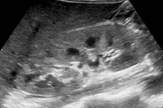
Diffuse changes in renal parenchyma and sinuses: what is it?
Last reviewed: 12.07.2025

All iLive content is medically reviewed or fact checked to ensure as much factual accuracy as possible.
We have strict sourcing guidelines and only link to reputable media sites, academic research institutions and, whenever possible, medically peer reviewed studies. Note that the numbers in parentheses ([1], [2], etc.) are clickable links to these studies.
If you feel that any of our content is inaccurate, out-of-date, or otherwise questionable, please select it and press Ctrl + Enter.
When explaining to patients what diffuse changes in the kidneys revealed during their ultrasound scan (US) mean, nephrologists and urologists talk about echographically visualized pathological deviations in the tissues and individual structures of this organ. Such pathomorphological signs are present in many kidney diseases, as well as diseases that give nephrological complications.
Therefore, the information obtained is very important for correct diagnosis and adequate treatment. [ 1 ]
Causes diffuse renal changes
The key causes of diffuse changes in the renal parenchyma, interstitial tissue of their stroma, cortex and medullary (medulla) substance, sinuses, Malpighian pyramids or tubular (canalicular) structures of nephrons are in the vast majority of cases associated with nephrological diseases:
- inflammation of the kidneys – nephritis (interstitial, chronic tubulointerstitial, diffuse fibroplastic, lupus, sarcoidosis or radiation);
- chronic pyelonephritis, including xanthogranulomatous;
- inflammation of the renal glomeruli - glomerulonephritis, which has several morphological varieties;
- glomerulosclerosis (with the formation of scars in the renal glomeruli);
- nephropathies – ischemic, obstructive, urate, IgA nephropathy (Berger's disease);
- cystic kidney diseases;
- kidney stone disease (nephrolithiasis);
- kidney damage in metabolic diseases, in particular, nephrocalcinosis - the deposition of crystalline calcium in the renal parenchyma and tubules with hypercalcemia or calcium oxalate crystalluria;
- renal neoplasms (lipoma, adenoma, adenocarcinoma, hypernephroma, sarcoma).
Similar changes are observed in tuberculosis of the kidneys, in arterial hypertension (leading to renovascular hypertension and nephrosclerosis), in diabetic nephropathy.
Diffuse changes in the kidneys in children revealed by ultrasound are also the result of pathological processes caused by nephrological, metabolic or autoimmune diseases. More details in the publications:
- Main kidney diseases in children
- Hereditary nephritis (Alport syndrome) in children
- Chronic glomerulonephritis in children
- Hereditary and metabolic nephropathy in children
In cases of such congenital anomalies of the structure of the renal parenchyma as hereditary polycystic kidney disease (multicystic renal dysplasia) and spongy kidney, in neonatal and infantile hydronephrosis, in diffuse nephroblastomatosis, as well as in hereditary tubulopathies (lesions of the renal tubules), ultrasonography can show existing diffuse changes in the kidneys of a newborn child. [ 2 ]
Risk factors
Factors that may increase the risk of chronic kidney disease with diffuse changes in kidney function include:
- hereditary predisposition (presence of nephrological diseases in the family history);
- diabetes;
- high blood pressure;
- rheumatoid arthritis;
- gout and nitrogen metabolism disorders;
- systemic lupus erythematosus (SLE);
- systemic scleroderma;
- myeloma disease;
- autoimmune hepatitis;
- protein metabolism disorders – amyloidosis (types AA and AL);
- hemorrhagic vasculitis;
- hyperparathyroidism;
- tuberculosis;
- kidney injuries with damage to their structure;
- invasion by echinococcus larvae;
- long-term use of certain medications (painkillers, antibacterial drugs, non-steroidal anti-inflammatory drugs, diuretics);
- immune deficiency;
- smoking, alcoholism, drug addiction.
Pathogenesis
Such a pathomorphological sign as diffuse changes in the kidneys is recorded by sonography as a fact indicating disturbances that cause the pathogenesis of specific diseases.
For example, in many types of nephritis or pyelonephritis, the mechanism of the appearance of diffuse changes consists of infiltration of parenchyma cells and interstitial tissue of the kidneys by leukocyte monocytes and tissue macrophages (maximally active during inflammation) and subsequent alteration in the form of focal or continuous transformation of functional cells into fibrous tissue. Also see – Pathogenesis of interstitial nephritis
Patients with lupus develop nephritis with damage to functional structures and segmental fibrosis of kidney tissue due to the effect of human antiphospholipid immunoglobulins (autoantibodies) on cell membranes and basement membranes of the walls of renal capillaries. [ 3 ]
Dysfunction of kidney nephrons in glomerulosclerosis is the result of worsening hypertrophy of the filtering glomeruli.
In the complex biochemical mechanism of diabetes-induced kidney damage, which, according to clinical statistics, occurs in a third of patients, the main role is given to elevated blood sugar levels (hyperglycemia). Under the influence of excess glucose, post-translational modification of membrane proteins of kidney cells is activated, which undergo glycosylation with disruption of their structure. And the simultaneous hyperactivity of protein kinase enzymes, affecting cellular metabolism, increases the toxicity of free radicals.
Diffuse changes in the liver, pancreas and kidneys in amyloidosis are explained by extracellular fibrous deposits of insoluble amyloid protein in the tissues of these organs. In the kidneys, this protein accumulates in the filtering structures of the nephrons, which causes nephrotic syndrome.
See also - Pathogenesis of chronic renal failure
Symptoms diffuse renal changes
Can patients experience symptoms of diffuse kidney changes – as characteristic manifestations of the disease or its first signs?
As noted above, these pathological changes are detected during instrumental diagnostics - during ultrasound examination of the kidneys and ureters, representing diagnostic indicators of a wide range of nephrological diseases or renal complications of other diseases. Of course, diffuse changes in the structure of both kidneys, changes in the left or right kidney - depending on the stage of the pathological process and its severity - significantly reduce the performance of the organ. But this is manifested by the symptoms of a specific disease. Read more:
Forms
According to the degree of expression on the ultrasound image, chronic diffuse changes in the kidneys, affecting the entire organ or part of it, can be weakly expressed or unclear; moderate and pronounced diffuse changes in the parenchyma and other structures are also determined.
According to nephrologists, diagnostic ultrasound often does not provide a specific result due to the lack of specificity of hyperechoic images, so clinical correlation is needed for its interpretation. What types of diffuse changes can be visualized in specific kidney diseases?
Diffuse changes in the parenchyma of both kidneys - with an increase or decrease in its thickness - can occur with inflammation and the development of urolithiasis; with nephrosclerosis and problems with the intrarenal vessels; with congenital anomalies, systemic, metabolic and immune-mediated diseases.
In cases of xanthogranulomatous pyelonephritis, renal parenchymal malakoplakia or lymphoma, there may be unilateral diffuse changes in the left kidney or only the right.
Diffuse changes in the renal sinuses (renal sinuses with the system of calyces and pelvises located in them) may indicate pyelitis and pyelonephritis (when the tissues of the sinuses become denser due to inflammation), as well as hydronephrosis (with dilation of the urine-filled calyceal-pelvic structures). In fibrolipomatosis in the sinus area, in the presence of cysts, stones or tumors, diffuse changes affect both the calyceal-pelvic system and the renal parenchyma. [ 4 ]
Diffuse changes in the renal stroma (formed by connective tissue) are most often detected in cases of its lymphoid and macrophage infiltration in acute and chronic inflammatory diseases (interstitial and tubulointerstitial nephritis, pyelonephritis), in patients with metabolic disorders or due to exposure to viral or exogenous chemical toxins. [ 5 ]
Diffuse-focal changes in the kidney are characteristic of nephrosclerosis, which develops as a result of impaired urine outflow, as well as chronic pyelonephritis with cicatricial tissue alteration and multiple foci of atrophy.
Diffusely heterogeneous changes in the kidneys with an increase in their size may be related to polycystic disease and amyloidosis, and localization of such changes in the parenchyma is possible with malignant renal tumors.
Diffuse non-specific changes in the kidneys often accompany their infiltrative lesions (cell infiltration) of inflammatory or tumor etiology. Such changes (in epithelial neoplasms of the renal parenchyma, renal cell or medullary cancer, sarcoma, lymphoproliferative diseases) can enlarge the kidney, but do not have a clear boundary between the lesion and the normal parenchyma. [ 6 ]
Complications and consequences
The internal capacity of the kidneys is limited, since the formation of new nephrons is impossible. Potential complications and consequences of diseases in which diffuse changes in the structures and tissues of the kidneys occur may manifest themselves:
- fluid retention in the body and tissue edema;
- hyperkalemia (increased potassium levels in the blood), which affects the functioning of the cardiovascular system;
- impaired tubular reabsorption of electrolytes;
- development of acute and chronic nephrotic syndrome;
- progressive decrease in glomerular filtration rate and deterioration of renal function;
- uremia and chronic renal failure requiring peritoneal dialysis;
- uremic coma;
- irreversible kidney damage – up to the terminal stage. [ 7 ]
Diagnostics diffuse renal changes
Ultrasound examination is the most important and quite informative part of kidney examination. Changes in their tissues and structures of a diffuse nature are visualized on the ultrasound monitor in different ways: in the form of anaechogenic, echo-positive, hypo or hyperechogenic formations, areas and regions. [ 8 ]
The echo signs of diffuse changes in the kidneys assessed by ultrasound diagnostic specialists are described using medical terminology and are intended for doctors, not patients. More information in the articles:
However, it is impossible to determine the cause of diffuse changes in the kidneys based on the results of ultrasound examination. Therefore, a full diagnosis is carried out – taking into account the patient’s medical history and complaints, using all currently available methods. [ 9 ]
First of all, laboratory tests are necessary: blood tests for creatinine and urea nitrogen levels, for antibodies to the anti-phospholipase A2 receptor (PLA2R);
General and biochemical urine tests, daily urine analysis, urine protein, urine bacteria, urine concentration tests, etc.
If there is a suspicion of oncology and idiopathic nephrological diseases, a histological examination of a tissue sample is required, for which a puncture biopsy of the kidney is performed.
In addition to ultrasound, instrumental diagnostics includes:
- radiography;
- excretory urography with contrast agent;
- pyelography;
- scintigraphy;
- ultrasound Dopplerography of the kidneys;
- computed tomography of the kidneys;
- magnetic resonance imaging (MRI) of the kidneys.
Differential diagnosis
Many kidney diseases have non-specific (similar) symptoms, and only differential diagnostics – based on a complete examination of the patient and consultations with other specialists – makes it possible to make the correct diagnosis.
Who to contact?
Treatment diffuse renal changes
After identifying diffuse changes, determining their cause and determining an accurate diagnosis, complex treatment of the diagnosed diseases is prescribed:
Prevention
Experts recommend prevention of kidney disease by treating urinary tract infections, especially chronic ones, and ultrasound monitoring of patients with chronic nephrological pathologies.
Forecast
When diffuse changes in the kidneys are detected, the prognosis for their further condition may depend only on the course of the diseases that caused them, many of which quickly progress to functional renal failure, and some, with successful therapy, are capable of partial remission with relapses.