Heart rhythm, also known as heart rate, determines the sequence and frequency of heart muscle contractions, which allows blood flow throughout the body.
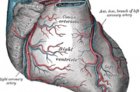
The coronary circulation (or coronary circulation) is the system of blood vessels that supplies blood and oxygen to the muscles of the heart, known as the myocardium.
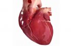
The heart is one of the most important organs of the complex system that is commonly called the human body. It is its engine, supplying blood to the most remote corners so that all organs receive sufficient nutrition and can function smoothly.
The tricuspid and pulmonary valves of the heart regulate blood flow from tissues to the lungs for oxygen enrichment, the mitral and aortic valves of the left heart control arterial blood flow to organs and tissues. The aortic and pulmonary valves are the outlet valves of the left and right ventricles, respectively.
The anatomy of the aortic valve is considered the most studied, since it was described long ago, starting with Leonardo da Vinci (1513) and Valsalva (1740), and repeatedly, especially during the second half of the 20th century.
It was previously believed that all heart valves were simple structures whose contribution to unidirectional blood flow was simply passive movement in response to an applied pressure gradient.
The tricuspid valve, like the mitral valve, consists of a complex of anatomical structures, including the fibrous ring, valves, tendinous chordae, papillary muscles and adjacent parts of the right atrium and ventricle.
The mitral valve is an anatomical and functional structure of the heart of a funnel shape, consisting of a fibrous ring, cusps with chords, papillary muscles, functionally connected with the adjacent parts of the left atrium and ventricle.
The pulmonary valve is separated from the fibrous framework of the heart by the muscular septum of the right ventricular outlet. This valve has no fibrous support. Its semilunar base rests on the myocardium of the right ventricular outlet.