Medical expert of the article

New publications
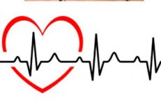
Heart rate
Last reviewed: 29.06.2025

All iLive content is medically reviewed or fact checked to ensure as much factual accuracy as possible.
We have strict sourcing guidelines and only link to reputable media sites, academic research institutions and, whenever possible, medically peer reviewed studies. Note that the numbers in parentheses ([1], [2], etc.) are clickable links to these studies.
If you feel that any of our content is inaccurate, out-of-date, or otherwise questionable, please select it and press Ctrl + Enter.
The rhythm of the heart, also known as the heart rate, determines the sequence and frequency of contractions of the heart muscle, which allows blood flow throughout the body. The human heart usually beats regularly and at a certain frequency.
There are several types of heart rhythm, including:
- Sinus rhythm: This is a normal heart rhythm in which contractions begin in the sinus node, which is located in the right atrium. This rhythm is considered normal and healthy.
- Arrhythmia: An arrhythmia is an unusual heart rhythm that may include a heartbeat that is too fast, too slow, or irregular. Examples of arrhythmias include atrial fibrillation (atrial fibrillation) and ventricular fibrillation (ventricular fibrillation).
- Tachycardia: This is a condition in which the heart beats too fast, often more than 100 beats per minute. Tachycardia can be caused by stress, physical activity, fever, or medical conditions.
- Bradycardia: Bradycardia is a condition in which the heart beats too slowly, less than 60 beats per minute. This may be normal for some athletes, but can also be a sign of heart problems.
- Extrasystoles: Extrasystoles are extra contractions of the heart that occur outside the normal rhythm. They can be ventricular or atrial. In most cases, they do not pose a health risk, but in some situations they may require medical attention.
- Avrent rhythms: These rhythms occur when heart signals follow unusual pathways in the heart, which can cause arrhythmias.
- Atrial flutter and atrial flutter: These arrhythmias are characterized by rapid and regular atrial contractions and may require treatment.
The rhythm of the heart can be assessed by an electrocardiogram (ECG), which records the electrical activity of the heart and allows physicians to determine whether the heart rhythm is normal or abnormal. A normal heart rhythm (sinus rhythm) has a rate that depends on the age and physiologic characteristics of the patient and is usually between 60 and 100 beats per minute.
Any changes in heart rhythm can be signs of heart problems, and doctors use ECG analysis and other techniques to diagnose and treat arrhythmias and other rhythm disorders.
Nervous regulation of heart rhythm
It is carried out by nerve signals that control the activity of the heart muscle. The main nerves involved in the regulation of heart rhythm are:
- Sinus node (sinoatrial node, SA node): This node is located at the top of the right atrium and serves as the "supplier" of the main impulse to the heart. It generates electrical impulses that initiate atrial contraction and thus the beginning of the cardiac cycle. The CA node is part of the automatic nervous system of the heart.
- Autonomic Nervous System: The autonomic nervous system (ANS) consists of sympathetic and parasympathetic subsystems. The sympathetic nervous system activates the heart, increasing heart rate and force of contraction. Conversely, the parasympathetic nervous system slows the heart rate and decreases the force of contractions. These two subsystems balance each other and regulate the heart rhythm according to the body's needs.
- Autonomic Nervous System: The autonomic nervous system (also called the autonomic nervous system) controls many of the body's automatic functions, including cardiac function. It includes the sympathetic and parasympathetic nervous systems and regulates the activity of the CA node and other parts of the heart.
Autonomic regulation of heart rate refers to the control of cardiac activity by the autonomic nervous system. The autonomic nervous system consists of sympathetic and parasympathetic subsystems that work in tandem to regulate various body functions, including cardiac function.
- Sympathetic Nervous System: Stimulation of the sympathetic nervous system activates cardiac activity. This occurs by increasing the heart rate and increasing the force of heart contraction. Sympathetic activation usually occurs in response to stress, physical activity, or other situations where the body needs to increase blood flow and adapt to stress.
- Parasympathetic nervous system: The parasympathetic nervous system, on the contrary, slows down cardiac activity. This occurs by increasing the time between heartbeats and decreasing the force of heart contraction. Parasympathetic activation usually occurs in states of rest and relaxation, when the body does not need a high rate of heartbeat.
Heart rate regulation is accomplished by central and peripheral mechanisms, including autonomic nerves, chemical signals, and hormones. The system of autonomic regulation of heart rhythm allows the body to maintain an optimal level of blood supply depending on current physiological needs.
- Central Nervous System: The hypothalamus and brain stem also play an important role in regulating heart rate by sending signals along nerve fibers to the heart via the autonomic nervous system.
This complex systems approach to heart rate regulation allows the body to adapt to different conditions and demands, maintaining optimal blood circulation and ensuring vital organ and tissue functions.
Heart rhythm norms by age
The following are general recommendations for normal heart rate (pulse rate) based on age:
- Newborns (0-3 months): 100-150 beats per minute.
- Infants (3-12 months): 90-120 beats per minute.
- Children (1-10 years old): 70-120 beats per minute.
- Adolescents and adults (over 10 years of age): 60-100 beats per minute.
These are general guidelines, and normal heart rate may vary slightly from person to person. It can also vary depending on physical activity, emotional state, and other factors. Athletes and very physically fit people usually have a lower resting heart rate.
Sinus rhythm of heartbeats
This is a normal heart rhythm in which contractions begin in the sinus node, which is located in the right atrium of the heart. The sinus node generates an electrical impulse that travels throughout the heart and causes it to contract. This rhythm is considered normal and healthy.
Sinus rhythm is characterized by the following features:
- Regularity: The intervals between heart contractions in sinus rhythm are usually regular.
- Frequency: The normal heart rate for adults is usually between 60 and 100 beats per minute.
- Normal waves: An electrocardiogram (ECG) in sinus rhythm usually shows characteristic P, QRS, and T waves that reflect the different phases of the cardiac cycle.
Sinus rhythm is considered the gold standard of normal cardiac activity and is the baseline for comparison of other rhythms and arrhythmias. It can change in response to physical activity, stress, posture changes, or other physiologic and pathologic factors. If sinus rhythm is inappropriately fast (tachycardia) or slow (bradycardia), it may be a sign of a medical problem and may require further evaluation and treatment by a physician or cardiologist.
Heart Rate Variability (HRV, Heart Rate Variability)
It is a measure of the variability of time intervals between consecutive cardiac contractions. This parameter makes it possible to assess the adaptive capabilities of the cardiac system and its response to various physiological and psychological factors. Heart rate variability is considered an important indicator of the state of the autonomic nervous system and can be used for medical and research purposes. Here are some key aspects of heart rate variability:
- AutonomicNervous System: Heart rate variability is related to the functioning of the autonomic nervous system, which controls the internal organs, including the heart. The autonomic nervous system includes sympathetic (stress) and parasympathetic (relaxation) branches, and heart rate variability reflects the balance between the two.
- Physiologic factors: Factors such as breathing, physical activity, food, and stress levels can influence heart rate variability. For example, deep and slow breathing is often associated with high heart rate variability.
- Heart Health: Research suggests that the level of heart rate variability may be related to heart health and risk of cardiovascular disease. A decrease in variability may indicate poorer heart health.
- Stress and emotions: Em otional states such as stress, anxiety and depression can affect heart rate variability. Increased stress is often accompanied by a decrease in variability.
- Methods of measurement: Heart rate variability can be measured using electrocardiography (ECG) or specialized devices such as pulse oximeters. There are various parameters and techniques for calculating variability, including time and frequency analyses.
- Clinical use: Heart rate variability is used in medical practice to assess patients with heart disease, diabetes, neurological disorders and other conditions. It can also be an indicator of risk of complications.
Heart rate variability can be a useful tool for both medical professionals and people who want to monitor their health and stress reactions. However, interpreting variability data requires some knowledge and experience, and therefore it is recommended to discuss the results with a qualified physician.
Normal heart rate variability (HRV) values can vary depending on many factors, including age, gender, physical activity, and health status. It is also important to note that there are several parameters and techniques for measuring HRV, and each may have their own normal ranges. However, general ideas about normal HRV values include the following:
- Total variability: The level of HRV total variability can be expressed as a number representing the average of the time intervals between heartbeats (R-R intervals) in milliseconds. Normal values can range from 20 to 100 milliseconds.
- Parameters in the frequency domain: HRV can also be measured using frequency analysis, which divides variability into different frequency components such as high frequency (HF) and low frequency (LF) bands. Normal values can vary depending on age and technique, but typically:
- HF (high frequency range) is normally between 20 and 100 ms².
- LF (low frequency range) is normally between 40 and 150 ms².
- The LF/HF ratio can vary, but low values may indicate a dominant influence of the parasympathetic (relaxing) nervous system.
- Diurnal variability: Heart rate variability can vary at different times of the day. It is common to see HRV increase during sleep and decrease during stress or activity.
It is important to remember that HRV is an individual parameter and normal values may vary from person to person. Therefore, it is important to have a careful measurement and interpretation in collaboration with a qualified medical professional to assess your heart rate variability and its value.
Assessment of heart rate variability
It is a method of studying the variability of the intervals between successive heartbeats (RR intervals) over time. This variability reflects the regulatory mechanisms of heart rhythm and may warn of abnormalities in them. HRV assessment can be useful in clinical medicine and research to evaluate the state of the autonomic nervous system and other physiologic processes. Here are some of the main aspects of HRV assessment:
- Measurement: HRV assessment is based on an electrocardiogram (ECG or EKG) recording that records the electrical activity of the heart over time. Using specialized software, the intervals between successive heartbeats are analyzed.
- RR intervals: HRV measurement assesses changes in the duration of RR intervals (intervals between heartbeats) over time. The intervals may be short or long, and their variability may contain information about the balance between the sympathetic and parasympathetic nervous system, as well as other factors.
- Analysis: There are several methods for analyzing HRV, including temporal and frequency methods. Temporal methods estimate statistical parameters of RR intervals, such as mean, standard deviation, etc. Frequency methods decompose HRV into different frequency components (e.g., high-frequency and low-frequency bands), which can provide information about the influence of the autonomic nervous system on heart rate.
- Clinical applications: HRV assessment can be useful in assessing the risk of cardiovascular disease, stress, depression and other conditions. It can also be used to monitor the effectiveness of treatment and training in athletes.
Assessing heart rate variability requires specialized equipment and software, as well as expertise to interpret the results. Therefore, if you are interested in HRV or need its assessment for medical purposes, you should consult a qualified physician or cardiology specialist.
Heart rhythm disturbance
A heart rhythm disorder, known as arrhythmia, is an alteration of the normal heart rhythm. Instead of regular and coordinated heartbeats, there are abnormalities in the frequency, regularity or sequence of heartbeats. Arrhythmias can be temporary and asymptomatic, but can also cause serious heart problems and require treatment. Here are some of the most common types of arrhythmias:
- Atrial fibrillation (AF): This is one of the most common arrhythmias. During AF, the atria begin to contract uncontrollably, creating erratic electrical impulses. This can lead to irregular ventricular contractions and increase the risk of blood clots and stroke.
- Tachycardia: This is an arrhythmia in which the heart beats too fast (more than 100 beats per minute at rest). Tachycardias can be sinus (normal) or caused by other mechanisms.
- Bradycardia: This is an arrhythmia in which the heart beats too slowly (less than 60 beats per minute at rest). It may be caused by problems with the sinus node (normal pedicle) or the anterior conducting system.
- Extrasystoles: Extrasystoles are extra heartbeats that can occur between normal contractions. They can be atrial or ventricular and are usually not a serious problem, but in rare cases can cause chest pain or discomfort.
- Blockages: Blockages are problems with the conduction of electrical impulses in the heart. They can be incomplete (partial) or complete and can affect normal ventricular contraction.
- WPW syndrome: This is an abnormality of electrical impulse conduction in the heart in which there is an extra pathway for impulses to be transmitted between the atria and ventricles. It can cause cardiac arrhythmias.
Increased heart rate
Heart palpitations, also known as tachycardia, is a condition in which the heart beats too fast, more often than the normal heart rate for the patient's age and condition. The normal heart rate for adults is between 60 and 100 beats per minute at rest.
Tachycardia can occur for a variety of reasons, including:
- Physical activity: The heart's normal response to physical activity is to increase heart rate to provide adequate blood supply to the muscles.
- Stress and anxiety: Strong emotional distress can cause palpitations.
- Heat and dehydration: Increased ambient temperature or insufficient fluid intake can cause tachycardia.
- Anemia: A lack of red blood cells and oxygen in the blood can cause palpitations.
- Hyperthyroidism (increased thyroid function): Elevated thyroid hormone levels can cause tachycardia.
- Medications and drugs: Some medications, such as adrenergic agents, can cause palpitations as a side effect.
- Cardiac arrhythmias: Uncontrolled arrhythmias can lead to tachycardia.
- Other medical conditions: Some medical conditions, such as infections and inflammation, can cause palpitations.
Tachycardia can be temporary and intermittent or become chronic. Chronic tachycardia may require treatment, especially if it is associated with cardiac arrhythmias or other heart conditions.
Slow heart rate
A slow heart rate (bradycardia) is a condition in which the heart beats slower than normal. The normal adult heart rate is usually between 60 and 100 beats per minute at rest. If your heart rate falls below this range, it may be a sign of bradycardia.
Bradycardia can be temporary or chronic and have different causes:
- Physiologic bradycardia: In some people, a lower resting heart rate is normal, especially in athletes and very physically fit people. This is called physiologic bradycardia.
- Autonomic Nervous System: Heart rate regulation is carried out by the autonomic nervous system. Perturbations in this system can cause bradycardia.
- Medications: Some medications, such as beta-blockers and some blood pressure medications, can slow your heart rate.
- Heart disease: Bradycardia may be associated with heart problems such as sinus node disease (the node that controls heart rhythm) or arteriosclerosis.
- Syncope: Some people may experience a slow heart rate periodically, which can cause fainting or syncope.
Bradycardia may be safe but may also require medical evaluation and treatment, especially if it is accompanied by symptoms such as dizziness, weakness, loss of consciousness, or chest pain.
Cardiac arrest
Cardiac arrest, also known as cardiac arrest (or asystole), is a critical condition in which the heart stops contracting and cannot provide blood flow through the body. This condition is extremely dangerous and requires immediate medical attention. Causes of cardiac arrest can vary and may include:
- Ventricular fibrillation (VFib): This is a serious heart rhythm disorder in which the ventricles begin to contract uncontrollably in an erratic manner. This can lead to complete cardiac arrest.
- Asystole: Complete absence of cardiac activity and electrical activity in the heart.
- Asphyxia: Asphyxiation or lack of oxygen in the body can cause cardiac arrest.
- Electrical failures: Electrical failures or blockages in the conduction of electrical impulses can lead to cardiac arrest.
- Severe allergic reactions: Anaphylactic shock caused by the allergen may cause cardiac arrest.
- Heart disease: Severe forms of heart disease, such as acute myocardial infarction, can lead to cardiac arrest.
It should be noted that cardiac arrest is considered a medical emergency requiring immediate resuscitation. If someone shows signs of cardiac arrest (e.g. Loss of consciousness, no pulse and no breathing), an ambulance should be called immediately and resuscitation (chest compressions and artificial ventilation) should be started. Resuscitation should be carried out by professional medical specialists, but anyone who is nearby can start the measures before the medical team arrives. A quick and correct response in such cases can save the patient's life.
Diagnostics of the heart rate
Heart rhythm diagnosis (ECG - electrocardiography) is the process of recording the electrical activity of the heart to assess its function and detect abnormalities. ECG is the standard method for analyzing heart rhythm and diagnosing various cardiac abnormalities. Here's how heart rhythm diagnosis works:
- Patient preparation: The patient is asked to undress to the waist to allow access to the chest. Electrodes are then placed on the skin of the chest, forearms and lower legs to record electrical signals from the heart.
- Performing an ECG: An electrocardiograph (ECG machine) records the electrical activity of the heart as a graph on paper or in electronic format. The process can last a few seconds to several minutes.
- ECG Interpretation: A cardiologist or ECG technician then analyzes the graph to determine the following parameters and characteristics:
- Heart rhythm: The physician determines whether the heart rhythm is normal (sinus rhythm) or abnormal (such as atrial fibrillation).
- Heart Rate: The average heart rate per minute (pulse) is determined to determine if the heart is in a normal rate range.
- R-R inter vals: The time intervals between heartbeats (R-R intervals) are analyzed to detect abnormalities.
- Abnormalchanges: The physician evaluates for abnormalities such as arrhythmias, blockages, enlargement of the heart chambers, and other changes.
- Additional tests: Depending on the ECG results and the patient's clinical presentation, additional tests such as Holter monitoring (continuous ECG recording for 24 hours), echocardiography (ultrasound of the heart) or stress tests may be ordered.
- Diagnosis and treatment: Based on the results of the heart rhythm diagnosis, the doctor will make a diagnosis and, if necessary, develop a treatment plan. Treatment may include drug therapy, procedures, or surgery.
It is important to note that heart rhythm diagnostics can be performed as part of a routine checkup or when symptoms related to heart problems such as chest pain, shortness of breath, severe fatigue, etc. Occur.
Holter heart rate monitoring
It is a diagnostic technique that continuously records the heart's activity (electrocardiogram or ECG) over a long period of time, usually 24 to 48 hours, sometimes even longer. This is called heart rhythm monitoring or Holter ECG. The name comes from Norman Holter, an American cardiologist who developed the first portable devices for such monitoring.
The purpose of Holter monitoring includes the following:
- Arrhythmia detection: This method can detect a variety of arrhythmias, including atrial fibrillation, ventricular fibrillation, extrasystoles, and other unusual heart rhythms that may be transient or not apparent during a standard ECG.
- Symptom Assessment: Patients who experience cardiac symptoms such as chest pain, shortness of breath, dizziness, or syncope (loss of consciousness) may wear a Holter monitor for a day or several days to record heart activity at the time of symptoms. This can help the doctor make a connection between symptoms and heart activity.
- Evaluating Treatment Effectiveness: If a patient is taking medications or undergoing procedures to treat arrhythmias, Holter monitoring can be used to evaluate the effectiveness of treatment and adjust medication dosage if necessary.
During monitoring, the patient wears a small, portable device that is connected to electrodes on the chest. The device records heart activity data throughout the period of wear, and the results are then analyzed by a physician.
Holter monitoring is an important tool for the diagnosis and management of arrhythmias and other cardiac conditions, especially those that occur suddenly or under certain conditions.
Pulsometer (or heart rate monitor)
It is a device that is used to measure your heart rate (pulse) and, in some cases, to monitor your heart rhythm. Pulsometers are widely used both for medical purposes and in sports training and fitness.
Basic heart rate monitor functions may include:
- Heart rate measurement: A heart rate monitor can measure your current heart rate, usually in beats per minute (bpm).
- Heart rate monitoring: Some advanced heart rate monitors can analyze the intervals between heartbeats (RR intervals) and provide information about heart rate variability. This is useful for assessing the state of the autonomic nervous system.
- Data Recording: Many heart rate monitors can record your heart rate data throughout your workout or day so you can analyze it later.
- Mobile connectivity: Some modern heart rate monitors can be linked to mobile apps via Bluetooth or other wireless technology, allowing you to track and analyze your performance on your smartphone or computer.
- Notifications: Some heart rate monitors can also provide notifications of calls, messages, and other events from your smartphone.
Heart rate monitors are available in a variety of forms, including wrist-worn devices, chest straps, smart watches and smart bracelets. Choosing a specific heart rate monitor depends on your needs and goals: for medical research, sports training or everyday health monitoring.
Who to contact?
Treatment of the heart rate
Cardiac rhythm restoration is the process of restoring a normal heart rhythm when arrhythmias or heart rhythm disturbances occur. Effective cardiac rhythm restoration can save lives in cases of cardiac arrest or serious arrhythmias. The ways to restore heart rhythm can vary depending on the situation and the patient's condition:
- Cardiopulmonary resuscitation (CPR): CRC is the primary method of restoring heart rhythm during cardiac arrest. It involves a series of chest compressions and artificial ventilation to maintain circulation and deliver oxygen to organs and tissues.
- Use of a defibrillator: Certain types of arrhythmias, such as ventricular fibrillation or atrial fibrillation, may require the use of a defibrillator. A defibrillator delivers a short electrical pulse that can help restore a normal heart rhythm.
- Medications: Doctors may use medicines to control and restore heart rhythm. For example, antiarrhythmic drugs may be used to manage certain arrhythmias.
- Cardioversion: This is a procedure that uses special equipment to establish a normal heart rhythm by delivering a controlled electrical shock through the chest.
- Electrophysiologic study and ablation: These procedures can be used to treat some cardiac arrhythmias, especially those that do not respond to medications or other methods.
Restoring heart rhythm is a complex and life-saving process that requires training and experience. In the event of cardiac arrest or serious arrhythmia, call for medical help and start CPR (if you know how to do it) and use a defibrillator, if available, until professional rescuers arrive. Quick action can save a life.
Restoring heart rhythm at home
It may be necessary if you or someone you love has heart rhythm problems such as atrial fibrillation (Atrial Fibrillation) or other arrhythmias. However, it is important to realize that restoring heart rhythm can be challenging and in some cases, medical attention is required. Here are some actions you can take at home depending on the situation:
- Atrial fibrillation (AF): Atrial fibrillation is a serious heart rhythm disorder in which the heart beats erratically and very fast. If you have diagnosed atrial fibrillation and have prescription medications, follow your doctor's recommendations and take your medications as prescribed. If serious symptoms occur (such as loss of consciousness), call an ambulance immediately.
- For other arrhythmias: If you notice symptoms of an arrhythmia, such as a feeling of heart palpitations, palpitations, or a slow heart rate, see your doctor for evaluation and diagnosis. Your doctor may prescribe treatments or procedures to normalize your heart rhythm.
- Helping others: If someone in your community has a serious heart rhythm problem, call an ambulance right away and follow the dispatcher's instructions for first aid until medical professionals arrive.
It is important to remember that heart rhythm intervention is a complex process that should be performed under the supervision of medical professionals. Listen carefully to and follow your doctor's recommendations and do not attempt heart rhythm restoration procedures without training and medical supervision.
Electrical pulse therapy for heart rhythm disorders
Electrical pulse therapy, also known as electrocardioversion or defibrillation, is a method of treating and restoring normal heart rhythm in certain cardiac abnormalities. This method is used to correct arrhythmias, especially atrial fibrillation (AF) and ventricular fibrillation (VFib), which can lead to cardiac arrest.
The principle of operation of electropulse therapy:
- Electrodes: The doctor applies special electrodes to the patient's chest. The electrodes are used to deliver an electrical impulse to the heart.
- Discharge: In PD or VFib, the heart may be involved in disordered electrical activity. Electrical pulse therapy uses a short, high-voltage electrical discharge (defibrillation) to "reset" the heart rhythm and return it to normal.
- Rhythm restoration: This is a procedure that allows the heart to start contracting again at a normal rhythm. If the procedure is successful, it can prevent the heart from stopping.
- Monitoring: Once the heart rhythm is restored to normal, the patient is usually monitored closely to ensure that the rhythm remains stable and there is no recurrence of arrhythmia.
Electrical pulse therapy is usually performed for heart rhythm abnormalities that are life threatening to the patient and cannot be treated with medication. It may be part of a medical emergency for cardiac arrest or to control arrhythmias in a hospital setting. The procedure is performed by professional medical specialists such as cardiologists or intensive care physicians.
It is important to realize that electro-pulse therapy is a serious medical procedure and it is only performed in strictly defined situations and under the supervision of experienced professionals.
Drugs for heart rhythm disorders
Medications for heart rhythm disorders, also known as antiarrhythmic drugs, are used to manage and control arrhythmias, including rapid heartbeat (tachycardia) and irregular heart rhythm. Your doctor will prescribe a specific medication depending on the type of arrhythmia, the patient's condition, and other factors. Some of the most common antiarrhythmic drugs include:
- Beta-adrenoblockers: These drugs reduce the activity of adrenaline, which can lower heart rate and reduce the risk of tachycardia. Examples include metoprolol, atenolol, and propranolol.
- Class I drugs (drugs that slow the conduction of an electrical impulse in the heart):
- Class IA drugs: Examples include kinidine, prokinamide.
- Class IB drugs: Examples include lidocaine, mexiletine.
- Class IC drugs: Examples include flecainide, propafenone.
- Class II drugs: These drugs also include beta-adrenoblockers, but they may be more specific for treating certain types of arrhythmias.
- Class III drugs: These drugs affect the duration of the action potential in the heart and can be used to treat different types of arrhythmias. Examples include amidarone, sotalol, dronedarone.
- Class IV drugs: These drugs are commonly used to control irregular heart rhythms and reduce heart rate. Examples include verapamil and diltiazem.
- Potassium antagonists: An example of such a drug is aminodarone, which can be used to treat a variety of arrhythmias.
- Other antiarrhythmic drugs: Depending on the individual case, your doctor may consider other antiarrhythmic drugs such as adenosine or ivabradine.
It is important to emphasize that the treatment of arrhythmias should be individualized, and the choice of a particular antiarrhythmic drug will depend on the diagnosis and characteristics of the patient. Medications can have side effects and their prescription should be made by the physician taking into account all risk and benefit factors. Patients receiving antiarrhythmic drugs should be monitored regularly by a physician to track the effectiveness of treatment and assess side effects.
Heart rhythm machines
Medical devices that help regulate heart rhythm, then the main ones are:
- Pacemaker: This is a medical device that is implanted in the patient's body, usually under the skin of the chest cavity, and is used to control the heart rhythm. The pacemaker generates electrical impulses to control the rate and rhythm of the heartbeat. It may be needed for bradycardia (slow heart rate) or other heart arrhythmias.
- Defibrillator: This is a device that is used to restore normal heart rhythm in the event of serious arrhythmias such as ventricular fibrillation or ventricular tachycardia. A defibrillator delivers a short electrical shock to reset the arrhythmia and return the heart to a normal rhythm.
- Implantable cardioverter defibrillator (ICD): This is a device that combines the functions of a pacemaker and defibrillator. It can be used in patients at high risk of developing serious arrhythmias and can automatically detect and correct them.
- External defibrillator: This is a portable medical device that is used in emergency situations to provide defibrillation. It is usually in automatic mode and can be used even by non-medical professionals if necessary.
These medical devices are used to treat cardiac arrhythmias and ensure a normal heart rhythm. Their use and implantation are performed by medical professionals, and patients who are prescribed them usually undergo a specialized medical examination and consultation to determine the best treatment method.