Endometrial biopsy
Last reviewed: 23.04.2024

All iLive content is medically reviewed or fact checked to ensure as much factual accuracy as possible.
We have strict sourcing guidelines and only link to reputable media sites, academic research institutions and, whenever possible, medically peer reviewed studies. Note that the numbers in parentheses ([1], [2], etc.) are clickable links to these studies.
If you feel that any of our content is inaccurate, out-of-date, or otherwise questionable, please select it and press Ctrl + Enter.

The study of the endometrium is based on the appearance of characteristic changes in the mucosa under the influence of steroid hormones of the ovary. Estrogens cause proliferation, and progesterone - secretory transformation. The study of the endometrium helps diagnose latent tuberculosis, determine the state of the uterine cavity and its walls.
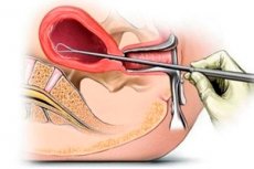
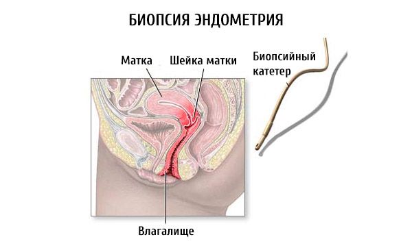
The material for analysis is obtained most often by the scraping method , which should be possibly complete, which also gives a therapeutic effect (for example, in dysfunctional uterine bleeding ). The vacuum aspiration method proved to be less traumatic and gives good results. The material is taken on the 21-24th day of the cycle, with acyclic bleeding at the beginning of the cycle, when the endometrium is preserved.
When evaluating histological preparations, the morphological features of the functional layer of the endometrium, the nature of the structure of the stroma and glands, as well as the features of the glandular epithelium are taken into account.
Normally, during the phase of secretion, the glands are enlarged, have a sawlike shape, compact and spongy layers are visible. The cytoplasm in the cells of the glandular epithelium is light, the nucleus is pale. In the lumen of the glands a secret is visible. With hypofunction of the yellow body, the endometrial glands are slightly convoluted, with narrow lumens.
In the anovulatory menstrual cycle, the endometrial glands are narrow or somewhat enlarged, straight or convoluted. The glandular epithelium is cylindrical, high: the nuclei are large, located basally or at different levels.

Atrophic endometrium is characterized by a predominance of the stroma, sometimes single glands are visible. Sam scraping is extremely meager.
The glandular cystic endometrial hyperplasia is characterized by cystic-enlarged glands, increased epithelial proliferation, often it is multi-nuclear, with thickened or cubic cells, the nuclei are at different levels.
Endometrial biopsy has great diagnostic value in assessing ovarian function . Secretory endometrium, removed when scraping endometrium 2-3 days prior to the onset of menstruation, indicates the occurred ovulation with an accuracy of 92%.
Who to contact?


 [
[