Medical expert of the article

New publications
Diagnostic scraping of the uterine cavity walls
Last reviewed: 06.07.2025

All iLive content is medically reviewed or fact checked to ensure as much factual accuracy as possible.
We have strict sourcing guidelines and only link to reputable media sites, academic research institutions and, whenever possible, medically peer reviewed studies. Note that the numbers in parentheses ([1], [2], etc.) are clickable links to these studies.
If you feel that any of our content is inaccurate, out-of-date, or otherwise questionable, please select it and press Ctrl + Enter.
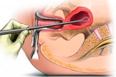
Diagnostic curettage of the uterine cavity walls is an instrumental removal of the functional layer of the uterine mucosa together with pathological formations that may originate from it. Diagnostic curettage of the uterine cavity walls should be performed only in a hospital setting with strict observance of the rules of asepsis and antisepsis. Anesthesia is provided by local paracervical anesthesia with 0.25% novocaine solution or mask anesthesia with nitrous oxide or intravenous anesthesia.
Diagnostic curettage of the uterine cavity walls is widely used in gynecological practice mainly to identify the condition of the endometrium. After probing the uterus, the cervical canal is dilated with Hegar dilators (usually up to No. 8). Then, the mucous membrane of the anterior and posterior walls of the uterus, its fundus and tubal angles is scraped with a medium curette.
If necessary, separate diagnostic curettage of the mucous membrane of the cervical canal and uterine cavity is performed.
The scrapings are sent separately for histological examination.
Indications for diagnostic curettage of the uterine cavity walls are uterine bleeding, dysfunctional menstrual cycle disorders, suspected malignant tumors of the uterus, placental and decidual polyps, hyperplasia and polyposis of the uterine mucosa, incomplete miscarriage, etc. In case of polyps, hyperplasia and incomplete miscarriages, curettage is performed not only for diagnostic purposes, but also for therapeutic purposes.
 [ 4 ]
[ 4 ]
After disinfection of the external genitalia and vagina, the cervix is exposed using mirrors, treated with alcohol and grasped by the anterior lip with bullet forceps. If the uterus is in retroflexion, it is better to grasp the cervix by the posterior lip. The uterine cavity is probed and the cervical canal is widened using Hegar dilators up to No. 9-10. The dilators are inserted, starting with small numbers, only with the force of the fingers of the hand, and not with the whole hand. The dilator is not brought to the bottom of the uterus, it is enough to pass it behind the internal os. Each dilator should be left in the canal for a few seconds; if the next dilator enters with great difficulty, the previous dilator should be inserted again. After widening the cervical canal, scraping of the walls of the uterine cavity begins, using sharp curettes of different sizes for this. The curette should be held freely, without resting on the handle. It is carefully inserted into the uterine cavity to the bottom of the uterus, then the curette handle is pressed so that its loop slides along the wall of the uterus, and it is brought out from top to bottom to the internal os. To scrape the posterior wall, without removing the curette from the uterine cavity, it is carefully turned 180°. Scraping is performed in a certain order: first, the anterior wall is scraped, then the left lateral, posterior, right lateral and corners of the uterus. The scraping is carefully collected in a jar with a 10% formalin solution and sent for histological examination.
There are certain features of uterine curettage that depend on the nature of the pathological process. An uneven, bumpy surface of the uterine cavity may be associated with interstitial or submucous myoma, so if this is detected, curettage should be performed carefully so as not to damage the capsule of the myomatous node. Damage to the capsule of the myomatous node may cause bleeding, node necrosis and its infection. The scraping may look like crumbly masses, characteristic of disintegrating malignant tumors. In such cases, complete curettage should not be performed so as not to perforate the uterine wall altered by the tumor. In all cases of suspected malignant tumor, separate diagnostic curettage of the uterine cavity should be performed.
Separate diagnostic curettage consists of first scraping the mucous membrane of the cervical canal, without going beyond the internal os. The scraping is collected in a separate test tube. Then the mucous membrane of the uterine cavity is scraped and this scraping is placed in another test tube. The directions for histological examination indicate from which part of the uterus the scraping was obtained.
After scraping the walls of the uterine cavity, the patient is taken to the ward on a gurney. Cold is applied to the lower abdomen. After 2 hours, she is allowed to get up. She is discharged under the supervision of a women's clinic on the 3rd day, if there are no complications and pain.