Medical expert of the article

New publications
Endometrial polyps in the uterus: causes of occurrence, signs
Last reviewed: 04.07.2025

All iLive content is medically reviewed or fact checked to ensure as much factual accuracy as possible.
We have strict sourcing guidelines and only link to reputable media sites, academic research institutions and, whenever possible, medically peer reviewed studies. Note that the numbers in parentheses ([1], [2], etc.) are clickable links to these studies.
If you feel that any of our content is inaccurate, out-of-date, or otherwise questionable, please select it and press Ctrl + Enter.
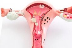
Polyps are neoplasms that are benign in origin. They form on the walls and in the uterine cavity and are a consequence of endometrial growth. They are usually attached to a special stalk. Polyps in the uterus are quite variable, ranging from miniature (1-2 mm) to large (3-4 cm). They are quite diverse, and can be multiple or single. The danger is that they are capable of quite intensive growth. Sometimes they extend far beyond the vagina. Absolutely all women are susceptible to polyp formation: from adolescence to post-menopause.
Is a polyp in the uterus dangerous?
It is dangerous in itself, and the complications it can cause are also dangerous. They often lead to infertility, hormonal imbalances and shifts. Hormonal secretion functions can be significantly disrupted. The greatest danger is that a polyp can develop into a malignant tumor and cause cervical cancer.
Less dangerous complications include the inability to conceive and the development of uterine bleeding. This is due to the fact that if left untreated and not removed in time, it develops its own vessels, which will gradually lead to it developing into a cancerous tumor. Increased risk of hemorrhage leads to the development of anemia, anemia, disruption of normal functioning, and a shift in the leukocyte formula. Constantly increasing. Large ones contribute to the appearance of increased bleeding, which is unable to stop on its own and requires surgical intervention.
In addition, the polyp is often subject to inflammation, an active infectious process develops, which spreads to the entire reproductive system. Foci of chronic inflammation are formed, since the polyp is unable to resist infection on its own.
Causes uterine polyps
The reasons why the uterus is subject to the formation of polyps remain completely unexplored. Most researchers believe that polyps are formed due to the fact that the body experiences a disruption of the hormonal background and nervous regulation. Numerous studies have established that growths have an increased sensitivity to hormonal substances, especially estrogenic ones. An increase in the content and activity of estrogen contributes to the increased growth of the neoplasm. A pattern has been established: the level of estrogen determines the rate of growth of the polyp. The higher it is, the larger the neoplasm will reach.
 [ 1 ]
[ 1 ]
Risk factors
The risk group includes women who are most susceptible to various endocrine disorders and have a genetic predisposition to dysfunctions of various glands. The risk of developing polyps increases in women who have a history of structural and functional disorders of the endocrine glands and are in a state of genetic restructuring of the body. The risk of developing polyps also increases significantly in women who are susceptible to stress and neuropsychic overstrain, who work a busy schedule, and do not observe the daily routine, rest, or work.
Women with various metabolic disorders also require careful attention: obesity, dystrophy, diabetes. The risk increases when taking a drug such as tamoxifen, which acts against breast cancer. But a polyp in the uterus may develop as a side effect. Women with high blood pressure and reduced immune status should also pay close attention to their health.
Pathogenesis
The pathogenesis is based on hormonal imbalance, which results in structural and functional disorders in the endothelium, the inner layer of the uterus. The mucous membrane is affected, and as a result of hormonal imbalances, it thickens. This leads to the formation of areas of increased density, in the place of which polyps are subsequently formed - neoplasms formed from thickened and overgrown mucous membrane. They tend to grow further and can spread throughout the uterus, multiply. Over time, they establish an independent system of blood supply, nutrition, and form their own genetic apparatus. This, according to most researchers, is the most dangerous when polyps appear. The establishment of independence entails transformation into one of the forms of cancer, uncontrolled growth and reproduction.
Symptoms uterine polyps
Often develop asymptomatically, without disturbing a person in any way. They appear when they have already reached a certain level of development and independence: at a later stage in the form of menstrual cycle disorders. Regularity changes significantly. There may be bleeding. Also, the occurrence of sudden bleeding during menopause, prolonged infertility, may indicate the development of polyps in the uterus.
The first signs that may indicate the development of polyps, albeit indirectly, are irregular menstruation, accompanied by bleeding. There is an increase in tissue, a gradual opening of the uterus. It tries to push the structure out. Intensive necrosis develops in it, blood circulation is disrupted. It manifests itself in painful and spastic sensations that can resemble light contractions.
Indirectly, long-term infertility or absence of menstruation in the absence of pregnancy indicates the formation of polyps. It is always necessary to remember that most polyps develop absolutely asymptomatically. They are often detected during examination. Therefore, the slightest sign that indicates a possible pathology or disorder should be a reason to contact a specialist.
If you have polyps in your uterus, you can have sex, as they do not affect sexual intercourse in any way. After removal, you should abstain from sex for at least a month.
Can a uterine polyp hurt?
In most cases, it develops asymptomatically and painlessly. But it can hurt when it has reached a large enough size, it divides, and multiple polyps form. Pain may indicate the beginning of transformation processes, in which the polyp begins to turn into cancer if the uterus tries to extract it. It opens up, pushes it out with contractions. This is accompanied by pain, since its innervation and blood circulation are disrupted. Twisting and squeezing of the stalk also causes painful sensations. This condition can be life-threatening and requires urgent surgical intervention.
[ 11 ]
Uterine polyp and pregnancy
If the size is small, the woman is able to conceive. But quite often such a pregnancy proceeds with complications. The main complication is considered to be placental abruption, which occurs as a result of the development of a polyp. This entails the threat of miscarriage or premature birth.
Another danger is that during pregnancy and childbirth, the polyp may be damaged. Damage always increases the risk of transformation into cancer.
But not everyone has such dire consequences. There are many known cases when polyps completely resolved during pregnancy on their own or under the influence of special therapy. Therefore, there is only one conclusion: each case is individual, and only the attending physician can predict how the pregnancy will proceed and how the polyp will behave in each specific situation, based on the results of tests and instrumental studies. For her part, a woman should be even more attentive to her health, and at the slightest concern or change, consult a doctor.
Is it possible to get pregnant with a polyp in the uterus?
Conception is possible. Especially when the polyp is small. But you need to understand that a polyp can become one of the causes of a complicated pregnancy, and many additional risks appear.
A somewhat different picture will be observed if the polyp is large and blocks the entrance to the uterus. This significantly reduces the likelihood of penetration of the fertilized egg into the uterus, complicates the possibility of its implantation, and increases the risk of developing an ectopic pregnancy.
[ 12 ], [ 13 ], [ 14 ], [ 15 ]
Polyps in the uterus after childbirth
If there was a small polyp before pregnancy, it may resolve during pregnancy or after childbirth. This is due to a decrease in the level of estrogen in the blood. It is during this period that the level of estrogen drops sharply, and a polyp, as is known, develops only with a high level of estrogen. A polyp can form after childbirth only after 2-3 months. Often, polyps occur after a complicated pregnancy, cesarean section, against the background of a general hormonal imbalance in the body.
Forms
There are many types of polyps. They can be single or multiple. According to the type of tissue and the nature of the lesion, glandular, fibrous, glandular-fibrous, adenomatous and placental are distinguished.
The endometrium is the inner layer of the uterus, its walls, cavity and mucous membrane. When polyps form, it becomes excessively compacted, as a result of which the compacted area gradually becomes isolated. Separate blood vessels are formed, the polyp acquires its own innervation. This contributes to its further growth. The danger of this type of polyps is that they can greatly increase in size. The membrane can swell so much that it gradually goes beyond the uterus and even fills the vagina.
Moreover, a polyp in the endometrium is capable of intensively dividing and forming multiple polyps that cover the entire uterine cavity. Such polyps require only scraping. Endometrial polyps are the most dangerous, since they have the highest risk of developing into a malignant tumor. They acquire an independent system of innervation and blood circulation, becoming relatively autonomous. Gradually degenerate into malignant neoplasms, acquiring the ability to divide uncontrollably.
Glandular polyp of the uterus
Formed in adolescence and youth. They are based on glandular cells. They are a cyst filled with fluid. They are a consequence of endometrial hyperplasia. These types of polyps are considered the most dangerous, since they have the highest risk of degeneration into a malignant tumor. In most cases, they cause bleeding, pose a threat during pregnancy and are often the cause of infertility. They grow very quickly and require surgical intervention.
Fibrous polyp in the uterus
Connective tissue underlies the formation of fibrous polyps. They are characterized by a fairly high density. They appear mainly before menopause and menopause, during hormonal changes.
Glandular fibrous polyp of the uterus
It is formed by individual elements of the endocrine glands, as well as connective tissue.
Adenomatous polyp of the uterus
Such growths are called adenomas. They contain altered cells and are often precursors to cancer. Such polyps quickly degenerate into cancerous tumors.
Polyp on the wall of the uterus
This type of polyp is a neoplasm that has arisen on the wall of the uterus from the mucous membrane. It is subject to active growth, since the mucous membrane is extensive, rich in blood vessels and nerves. There is a risk of developing a cancerous tumor. It can affect the ability to fertilize. This largely depends on its localization. If the polyp is located in the area where the implantation of the fertilized egg and further development of the placenta should occur, fertilization is impossible. In this case, as the polyp develops, its introduction into the mucous membrane, infertility may develop.
Polyp in the uterine cavity
In the uterine cavity, a polyp may be dangerous, or it may not cause any harm. Everything depends on the degree of its development and localization. If the polyp is large enough, it must be removed. If it is small, it is not necessary to remove it. The main thing is that it is not located in the cervix. A polyp located in the cavity can resolve on its own during pregnancy. This is due to a change in hormonal levels. The amount of estrogen decreases and the amount of progesterone increases.
A polyp in the uterine cavity during pregnancy must be constantly monitored, since it can twist, get damaged, which is fraught with serious consequences. Bleeding may occur, the risk of malignant degeneration of cells increases.
Endometrial polyp at the bottom of the uterus
A polyp located at the bottom of the uterus is considered the safest, since it is practically not exposed to external influences and mechanical damage. The danger is that it can grow to large sizes, and then surgical intervention will be required. If the polyp is small, it can resolve on its own with the help of conservative therapy.
Cervical polyps
Cervical polyps are the main cause of infertility and cervical cancer. Located in the cervix, the polyp blocks the fertilized egg from entering the uterus. As a result, its implantation into the mucous membrane and further development become impossible. The egg dies, or an ectopic pregnancy develops, which is a life-threatening condition for the woman.
There is also a high risk of developing a malignant tumor, since with such a location the polyp is mobile, well supplied with blood and nutrients. Gradually, it acquires its own blood vessels and transforms into a cancerous tumor. Also, in this place, the polyp is constantly exposed to mechanical damage, movements, which also increases the risk of malignant degeneration.
There is a risk of bleeding, since in such a place the uterus has a high reflex sensitivity and contractile activity. It perceives the polyp as a foreign body and begins to push it out into the vagina. This can lead to serious injury to the polyp and damage to blood vessels.
Cervical canal polyp
If a polyp occurs in the cervical canal, it must be removed as quickly as possible. Firstly, it will grow and block the lumen of the canal. Secondly, the probability of fertilization is reduced to a minimum as the polyp grows. The larger the polyp, the lower the chances of getting pregnant, since the egg cannot penetrate the uterine cavity. Even if you manage to get pregnant, the risk of complications increases sharply during childbirth. The pregnancy itself can also proceed with complications. This is due to the fact that the polyp is injured over time. During childbirth, it can be damaged, and even completely torn off, since it is located directly in the birth canal. This is dangerous due to bleeding, infection and inflammation.
Polyp of the uterine body
Develops directly in the body of the uterus. Formed as a result of proliferation of the endometrium. Usually such a polyp is held on a thin stalk, its size varies from several millimeters to several centimeters. Can cause heavy bleeding. Often cause infertility and cancer.
Diagnostics uterine polyps
It is important to undergo diagnostics in a timely manner. It is always necessary to remember that the pathology develops asymptomatically, without showing any signs. This is their danger. They can show themselves when it is already too late. Often they appear only when bleeding begins, anemia appears, or a malignant tumor develops. This indicates the importance of timely diagnostics and preventive examinations. Most polyps are detected during a routine examination.
Gynecological examination, laboratory and instrumental methods of research are used. During the examination, the doctor receives the necessary information about the structure and condition of the mucous membranes, about the morphology of the organs. Visually, it is possible to assess and suspect the presence of an inflammatory and infectious process, tumors. This is the basis for further assignment of the necessary tests, differential diagnosis, and establishment of a clinical picture.
An important role is given to ultrasound examination, during which the doctor evaluates the condition of the mucous membrane, the degree of its development. Hyperemia, swelling of the mucous membrane, and changes in thickness can be noticed. Changed echogenicity areas, visible on ultrasound, can indicate the presence of a malignant or benign tumor. It is also possible to detect bulges and areas of compacted mucous membrane, which can subsequently transform into tumors. This makes it possible to identify tumors and prerequisites for their formation at an early stage, and to take the necessary preventive and prophylactic measures in a timely manner.
Instrumental diagnostics
The main and most informative method for detecting polyps in the uterus is hysteroscopy. This is a method during which a hysteroscope is inserted into the body - a thin and fairly flexible device with a video camera at the end. Using this method, you can examine the walls of the uterus from the inside, the condition of the mucous membrane and detect a polyp if present. You can also take measurements, during which the doctor receives information about the size, volume and localization of the polyp. You can also count the number of polyps: single or multiple.
The great advantage of this method is that a biopsy can be taken during the procedure. During this examination, the doctor cuts off a small piece of mucous membrane for further histological examination. This is important when areas of increased density or neoplasms of unclear origin are detected. Histological analysis will show whether the tumor is benign or malignant.
Quite often, metrography is used. This method is one of the types of X-ray examination, during which a contrast agent is used, which is poured into the uterus. The image is then visualized. With the help of this method, it is easy to detect bulges, structural changes, as well as intracavitary changes in the uterus.
Diagnostic curettage can be used to obtain diagnostic information, during which accumulated material is removed from the uterus. It is then examined. Additionally, a hysteroscope is used, which makes it possible to examine the condition of the uterus from the inside. Sometimes there are cases when a hysteroscope is not used. In this case, the method is called blind curettage.
Biopsy of uterine polyps
The essence of a biopsy is that during the examination, biological material is collected using special instruments and methods. The material is then subjected to further histological analysis, which makes it possible to differentiate the tumor and make a final diagnosis. This method determines whether the tumor is benign or malignant.
Histology of uterine polyp
Histological examination is the study of a tissue sample obtained by biopsy using various biological methods. To perform a biopsy, a piece of the sample is taken and subjected to preliminary microscopy. For this, the preparation is stained using a convenient method that best suits the requirements and conditions of the laboratory, the type of microscopy used. During such a study, it is possible to detect changes in the cell that indicate various pathologies, such as cancer, inflammation, edema.
Having received such preliminary information, the laboratory technician develops further research tactics. The sample is seeded on a special selective medium intended for the growth of tissue cultures. The cultures are incubated in a thermostat at human body temperature. Whether the tissue grows on the nutrient medium determines whether the tumor is malignant or benign. A malignant, cancerous tumor grows intensively on the medium, while a benign tumor does not.
Then, based on the nature, direction and growth rate, the tumor species and its features are determined. It is possible to use the obtained information to further predict the tumor growth rate, select the optimal treatment method, evaluate the effectiveness and make the necessary adjustments.
Differential diagnosis
Differential diagnostics is based on precise differentiation of diagnoses. It is necessary to identify the signs of the disease that a person has and differentiate them from diseases that have similar features. For example, it is necessary to differentiate a common polyp from an atypical one, which can later transform into a malignant tumor. A biopsy will help to understand this.
It is also important to differentiate a polyp from malignant changes in the endometrium, or from malignant neoplasms such as sarcoma, carcinoma. Any type of cancer can only be excluded by biopsy and further histological examination. These are the most accurate methods, which are very informative.
It is important to differentiate a single polyp from multiple and growing ones, as they can cause infertility and severe bleeding. Hysteroscopy is used for this purpose.
Treatment uterine polyps
Treatment tactics depend on the results of analyses, histological and instrumental studies and are determined by the attending physician and other highly qualified specialists. Self-medication often ends in death.
Treatment of uterine polyps without surgery
Conservative treatment without surgery is possible, but it is not advisable. It is possible to cure with medications only when a single small polyp is detected. It can be significantly reduced or completely eliminated by special medications. If the patient is young or very young, it is worth trying medication therapy first. This is due to the high level of regeneration and recovery capabilities of the body. The immunity of a young girl is quite high and powerful and has all the necessary potential to overcome the disease on her own. Special medications are used that help increase immunity and resistance of the body. There is also special therapy aimed at suppressing the activity of the polyp, preventing its growth and reproduction. If there is a risk of developing a malignant tumor, special medications are used aimed at preventing the development of cancerous tumors.
Treatment includes hormonal therapy. Since the main trigger for polyp development is high estrogen levels and imbalance of other hormones, medications are taken that normalize the overall hormonal background and reduce estrogen levels. At the same time, progesterone levels increase, causing the polyp to shrink, atrophy, and be excreted during menstruation.
In the presence of polyps, women under 35 years of age need to take estrogen-gestagen contraceptives, which restore hormonal balance. The treatment regimen is selected by a doctor. Women over 35 years of age need to take drugs from the gestagen group, for example, duphaston, utrogestan. Women over 35 years of age should take releasing hormones, which prevent the undesirable effects of luteinizing hormones and estrogens.
In case of inflammation and infection, antibacterial drugs are additionally taken. Usually, antibiotic therapy is prescribed after preliminary bacteriological examination with determination of sensitivity to the isolated pathogen. This method makes it possible not only to isolate the main pathogen, but also to select the antibiotic to which it will show maximum sensitivity. The optimal dosage of this drug is also selected. In addition, folk methods are used, but they can only be used as part of complex therapy and after a preliminary consultation with a doctor.
If you have managed to completely get rid of the polyp, long-term observation and regular examination by a gynecologist are required, since polyps have the ability to self-repair and after some time, relapses of the disease may be observed.
Medicines
Medicines should be taken with caution and precautions. The main such measure is a preliminary consultation with a doctor and preliminary laboratory and instrumental control. Almost all drugs are selected in accordance with the test results, and after a preliminary check for effectiveness and compatibility. In some cases, even the dosage is selected in laboratory conditions. This is due to the fact that the drug must be selected as accurately as possible and have an idea of how the polyp may react to it. Any slightest incorrect action can lead to the degeneration of the cell from normal to cancerous, which will trigger the oncological process. In addition, the wrong selection of the drug, its dosage or method of administration can lead to severe bleeding, infertility.
If the development of a polyp is accompanied by pain (this most often happens if it has reached a large size, as well as when the stalk is twisted), you need to take painkillers, such as no-shpa. Take 50 mg 2-3 times a day.
Sometimes polyps may be accompanied by reddening of the mucous membrane, swelling, pain and itching in the perineum. After removal of polyps, postoperative swelling may also occur. In this case, it is recommended to take suprastin (150 mg 1-2 times a day).
If suprastin does not have any effect within 2 days, it is advisable to take a stronger drug - loratadine. Take 1 tablet per day, since it is a prolonged-action drug. The effectiveness lasts for 24 hours.
Often, against the background of changes in hormonal status, severe headaches and migraines develop. It is recommended to use pyrocetam, 1 tablet 2-3 times a day.
Vitamins
With polyps, the body needs vitamins. It is necessary to take vitamins both during treatment and in the recovery period after their removal. It is recommended to take vitamins in the following daily doses:
- Vitamin PP – 60 mg
- Vitamin H – 150 mcg
- Vitamin C – 500 mg
- Vitamin D – 45 mg
- Vitamin K – 360 mcg.
Read about treating uterine polyps with folk remedies in this article.
Hormones for uterine polyps
The main reason for their occurrence is hormonal imbalance. They develop when the level of estrogens increases sharply. Hormonal agents aimed at correcting the hormonal balance are often prescribed.
Preferably, progesterone preparations are prescribed. Progesterone and oxyprogesterone have proven themselves well. They are taken 1-2 times during the menstrual cycle, 125-250 mg, according to a specially selected regimen. The treatment regimen is selected by the attending physician for each patient individually. There are no general recommendations and regimens, since everything depends on the results of the studies.
Antiestrogenic complexes such as clomiphene are prescribed. Take 50 mg per day for 5 days. During the treatment, it is necessary to undergo ultrasound periodically, which will allow monitoring the likelihood of side effects.
Physiotherapy treatment
Physiotherapy is used for polyps. Various methods are used, but magnetotherapy, ultrasound therapy and electrophoresis have proven themselves to be the best. Magnetotherapy is used to resolve inflammatory processes, remove scars and postoperative sutures. Ultrasound can penetrate deep into tissues and affect them. It has a warming effect, resolves seals, restores damaged tissues, blood vessels, and prevents further development of polyps, their formation from the remaining tissues.
Electrophoresis is based on the effect of microcurrents on tissues. A bandage soaked in a medicinal product is applied to the mucous membranes or skin. The current affects the body, as a result of which the effect of the medicinal product is enhanced and its penetration into the tissues is ensured. Due to this, the drug penetrates deep into the tissues and has the necessary effect there. This significantly reduces the need for medicinal products and their dosage. Accordingly, the likelihood of complications and side effects is reduced.
Surgical treatment
Surgical treatment is based on the removal of polyps. This operation is also called polypectomy. The operation to remove a uterine polyp is quite simple. It can be performed conservatively, in which an open abdominal operation is performed. But today it is possible to perform it laparoscopically - through a special access using a laparoscope. Recovery after this method is slow, it is bloodless, the risk of complications is reduced to a minimum.
Methods of removing polyps in the uterus
There are three main methods by which they are removed: traditional, hysteroscopic, laparoscopic. The traditional method involves a regular abdominal operation, in which the polyp is removed. Almost no one uses this method anymore, it has long been outdated. It is dangerous due to numerous complications, recovery takes a long time, and the risk of bleeding increases. The operation is also quite complicated and lasts a very long time. The main difficulty is that it is necessary to consistently cut all the overlying layers, right down to the uterus itself, carry out the necessary manipulations to remove the polyp, and suture all the layers.
Hysteroscopy is used quite often. Until recently, this was the main method. It is considered low-traumatic, is performed under light anesthesia, and requires little time. During this time, all necessary manipulations are carried out, and the polyp is removed. If this method is used, recovery occurs quite quickly, and the patient can be discharged within 24 hours. The operation is performed by opening the cervix with special instruments. If multiple polyps are detected, curettage is performed under the control of a hysteroscope.
The most modern method is laparoscopic removal, which is performed using a special instrument - a laparoscope using a small laparoscopic access, which is made in the form of small incisions on the abdominal side (lower abdomen). The method is minimally invasive. Recovery is very fast, there are practically no scars. Postoperative pain also practically does not bother the patient. During the operation, an incision with a diameter of only 0.5-1.5 cm is made. If multiple polyps are detected or there is a high probability of developing uterine cancer, the uterus is removed.
Removal of polyp in the uterus
Most doctors try to use the laparoscopic method whenever possible, as it has a number of advantages over other methods. The main advantage is that the operation is performed not through a regular abdominal incision, but through laparoscopic access. With this method, small incisions are made in the lower abdomen. A special device, a laparoscope, is inserted through these incisions. The diameter of the incisions does not exceed 1.5 cm. This means that with this method there are practically no scars, the stitches heal quickly and recovery is also quite fast. The method allows you to avoid postoperative pain and blood loss. The risk of complications is reduced to a minimum.
After the incision is made, carbon dioxide is pumped into the uterine cavity. This allows the abdominal cavity walls to expand, eliminating the risk of damage to adjacent organs, and makes it easy for the surgeon to perform the necessary manipulations. The laparoscope is inserted into the cavity. At the end, it contains a small camera that visualizes the image on the screen.
The doctor examines the polyps in the uterus in detail and determines the tactics for further surgery. Using a laparoscope and other special equipment, the doctor excises the polyp and takes it out. After this, the cavity is examined again, the equipment is taken out. Sutures are applied. In just a few hours, the woman is transferred from the surgical department to the gynecological ward. The woman remains under observation for 5-7 days. In about 2 weeks, full recovery of working capacity occurs. The method is effective in cases where the risk of developing a malignant tumor is high, since the likelihood of metastases and tumors is practically excluded.
Prevention
Since the main cause of polyp development is ovarian dysfunction and excess estrogen, prevention comes down to preventing the development of ovarian dysfunction, as well as preventing endocrine disorders. To do this, it is necessary to regularly visit a gynecologist, endocrinologist, and undergo regular examinations.
It is important to eat healthy, home-cooked food. Avoid fast foods and genetically modified foods. It is necessary to maintain personal hygiene and hygiene of the genitals, and not to have promiscuous sex. It is also important to promptly treat chronic diseases and maintain immunity in a normal state. This is facilitated by walks in the fresh air, sports and physical exercise. You cannot overcool or sit on a cold surface.
Forecast
If uterine polyps are diagnosed in a timely manner and the necessary manipulations are performed to treat or remove them, the prognosis may be favorable. The prognosis may be unfavorable if no measures are taken or the doctor's recommendations are not followed. The most dangerous polyps are large ones and those that contain atypical cells in their structure. Such polyps may develop into a malignant tumor.