Medical expert of the article

New publications
Probing the uterine cavity
Last reviewed: 04.07.2025

All iLive content is medically reviewed or fact checked to ensure as much factual accuracy as possible.
We have strict sourcing guidelines and only link to reputable media sites, academic research institutions and, whenever possible, medically peer reviewed studies. Note that the numbers in parentheses ([1], [2], etc.) are clickable links to these studies.
If you feel that any of our content is inaccurate, out-of-date, or otherwise questionable, please select it and press Ctrl + Enter.
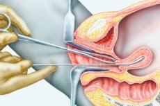
Uterine cavity probing is an operation to determine the direction of the uterine cavity, its length and the state of the wall relief. Uterine probing is performed with a uterine probe made of soft metal, 25 cm long, 3 mm in diameter. At the end of the probe there is a button and a thickening at a distance of 7 cm from the button, corresponding to the normal length of the uterine cavity; centimeter divisions are applied to the surface of the probe.
Probing of the uterine cavity is performed as a diagnostic procedure before an artificial abortion, and also to determine the length of the uterine cavity before diagnostic curettage. Probing has relative value for detecting submucous myomatous nodes.
The cervix is exposed with speculums. Its anterior lip is taken with bullet forceps and brought down. A probe is inserted through the cervical canal. It must be advanced carefully so as not to make a false move or perforate the uterine wall.
 [
[ Indications for the procedure
Probing is performed before diagnostic curettage of the uterine cavity, during abortion, to determine developmental anomalies of the uterus, submucous node in the uterus.
Technique uterine probing
First of all, the uterine probe is bent according to the position of the uterus determined during a bimanual vaginal examination. After disinfection of the external genitalia, the cervix is exposed using speculums, the vagina and the vaginal part of the cervix are wiped with alcohol. The anterior lip of the cervix is grasped with bullet forceps, after which the elevator is removed, and the speculum is handed over to the assistant to hold. With the left hand, the operator lowers and fixes the cervix with bullet forceps, and with the right hand takes the probe so that its handle lies freely between the thumb and index finger. The probe is inserted into the cervical canal and, without applying force, carefully advanced into the cavity to the bottom of the uterus. Upon completion of probing, the probe is removed, the bullet forceps are removed, and the vaginal part of the cervix is lubricated with iodine.
The length of the uterine cavity is determined by the scale of the uterine probe. An increase or decrease in its length indicates pathology ( adenomyosis, uterine myoma, uterine hypoplasia, etc.). Different lengths in the area of the uterine angles indicate its asymmetry. The direction of the probe is determined by the position of the uterus: in the anteflexio position, the probe is directed forward, in the retroflexio position - backward. The relief of the walls of the uterine cavity is normally smooth and even. A dense uneven surface protruding into the uterine cavity indicates the presence of a submucous myoma. Areas of soft consistency are suspicious of a malignant process. In case of uterine developmental anomalies, a uterine septum or double uterus is determined. Bloody discharge during or after probing may appear due to slight tissue injury, polyposis, endometritis, or uterine cancer.
Contraindications to the procedure
Contraindications for uterine probing are: acute and subacute inflammatory processes of the genital organs, III-IV degree of vaginal cleanliness, suspicion of uterine pregnancy.
Complications after the procedure
When probing the uterus, a false passage may be formed or its wall may be perforated. This may occur if a vaginal examination has not been performed before probing and the position of the uterus has not been determined, and also if the probe was inserted with force.