Medical expert of the article

New publications
Why have moles become bulging and what to do?
Last reviewed: 04.07.2025

All iLive content is medically reviewed or fact checked to ensure as much factual accuracy as possible.
We have strict sourcing guidelines and only link to reputable media sites, academic research institutions and, whenever possible, medically peer reviewed studies. Note that the numbers in parentheses ([1], [2], etc.) are clickable links to these studies.
If you feel that any of our content is inaccurate, out-of-date, or otherwise questionable, please select it and press Ctrl + Enter.
The appearance of moles on the body is a normal physiological process that has its own reasons. Small flat nevi on the baby's body are touching to parents. A cute mole on the cheek, shoulder or buttock is even considered a kind of charm or "highlight" in a person's image. Whatever the reason for the appearance of moles, they will always be the object of close attention, since information received from everywhere about the possibility of nevi degenerating into melanoma remains relevant in any case. Particular concern arises when we notice that moles have become convex, changed color or shape.
Causes convex mole
The fact that a mole has changed shape does not mean that skin cancer is developing in its place. Let's say that initially most moles are flat. The exceptions are angiomas and small convex moles that appeared in childhood. Moles themselves are not malignant neoplasms on the skin, and they tend to grow with a person, becoming larger in diameter and slightly rising above the surface of the skin.
However, this does not mean that this process should not be given importance. If a flat mole has become convex, but has not been injured and has not been subjected to excessive ultraviolet radiation (has not sunbathed or used a solarium), then the probability of its degeneration is very small. Its gradual growth and slight elevation above the skin surface will most likely be a safe natural process.
It's a different matter if the mole was subjected to mechanical impact or ultraviolet rays, after which its active growth was noticed. This is already a cause for concern and a reason to visit your dermatologist once again, and possibly a dermato-oncologist.
Pathogenesis
The thing is that moles are places where the body's defense is minimal. The so-called "weak spot" of a person. This is especially true for border nevi, such seemingly harmless dark, usually flat spots. The impact of negative factors on these areas, such as sunlight or injuries (blows, wounds, burns, strong friction, bites, etc.), leads to a change in melanocytes and their transformation into cancer cells, which, due to the weak connection between themselves, tend to quickly spread throughout the body in the form of metastases.
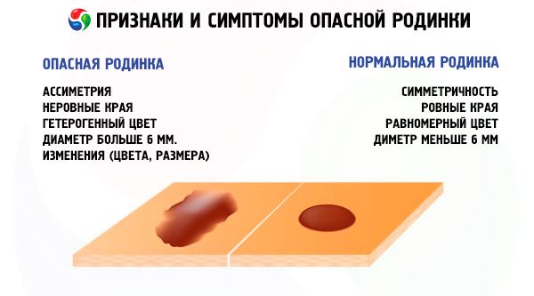
The fact that a flat mole has become convex, changed its size and shape, may be one of the symptoms of its degeneration into a malignant neoplasm. But we can talk about a high probability of this process only in the presence of other signs of degeneration, such as:
- Change in the color of a mole: the appearance of dark spots with signs of tissue death, uneven coloring, redness at the site of the nevus.
- Change in shape: noticeable asymmetry of the nevus, uneven edges, active growth.
- Unusual sensations: itching, tingling, burning or pain at the site of the nevus.
By and large, only a specialist can answer the question of why a mole has become convex based on a patient survey and certain studies on benignity. If you are worried about a mole, it is better to visit a dermatologist again and make sure that everything is fine with the mole than to start the process and reap the unpleasant fruits of your shortsightedness.
Complications and consequences
The consequences can be very tragic if the development of melanoma is not prevented in time. The initial stages of this insidious disease are easily treated. With a timely and correct approach to the treatment (removal) of a mole, recovery occurs quite quickly and without complications. If the cancerous tumor has taken root (metastasized) in other organs, the prognosis becomes far from rosy. Sometimes such carelessness can cost a person not only health, but also life.
Diagnostics convex mole
Any mole, including a convex one, is of particular interest to a doctor in terms of the possibility of its transformation from a benign formation into a cancerous tumor. It is worth understanding that whatever the reason for the appearance of a mole, whatever it consists of (melanocytes or vascular network), any changes in it indicate that a certain process has begun in certain layers of the skin. But whether the growth of a mole or its elevation above the skin is associated with the development of cancer can only be determined by a doctor using differential diagnostic methods.
It is only possible to determine by eye whether a mole can be dangerous, but only the results of instrumental examinations can tell how accurate such a diagnosis is. However, something can be learned from the patient's story, which will let the doctor understand whether such examinations are necessary.
When talking to a dermatologist, it is important to mention that the moles have become convex, although they were initially flush with the skin. You need to tell when the pigment spot appeared, whether it was exposed to traumatic factors, what sensations you experience in connection with the growth of the nevus. The doctor may ask questions about your family, about past generations, to find out the hereditary predisposition to the development of oncological diseases, in particular melanoma, and also inquire about your health.
Instrumental diagnostics of a convex mole is carried out using a special microscope with 10x magnification, which is called a dermatoscope. This device allows you to get a clear, detailed image of the nevus, which the doctor sees on the monitor screen, and also to create and save a diagram of moles on the human body in the computer memory for future diagnostic studies.
An even more complete picture is provided by one of the options for dermatoscopy - computer epiluminescent diagnostics, which allows for the illumination of a mole in depth.
If melanoma is suspected, a radioisotope study is sometimes prescribed, when a person is given a special preparation with radioactive phosphorus to drink on an empty stomach, and then the amount of the isotope in the mole and in the area of skin symmetrical with the pigment spot is measured.
The thermometric method of diagnosing nevi is based on different temperature readings of a healthy skin area and one affected by melanoma. This difference can reach 4 degrees.
Diagnosis of a convex mole does not include the usual tests for us. They may be required only in the case of surgical removal of the neoplasm. However, the most important role in the diagnosis of moles is given to such a type of laboratory research as a histological study of nevus cells. A biopsy is also a kind of analysis, but a piece of tissue from the mole that causes suspicion is taken for the study.
Most often, a biopsy is performed after the suspicious mole has been removed. Research of the material obtained as a result of the operation will help to understand how dangerous the neoplasm was and whether there were any metastases left behind.
A variant of histological examination without removing the mole is a puncture biopsy, when, using a special needle under local anesthesia, several cells of the nevus are taken for further examination.
Any type of histological examination gives a virtually 100% accurate diagnosis. If doubts still remain or there is no possibility to perform a biopsy, and everything indicates that there is a high risk of developing melanoma, the patient may be sent for an ultrasound, X-ray or even an MRI.
What do need to examine?
Who to contact?
Treatment convex mole
The most relevant type of treatment for moles is their removal. If moles have become convex, but studies have not confirmed their malignancy, the only indications for their removal may be an inconvenient location of the nevus, leading to trauma, or a hereditary predisposition to oncological diseases. Removal of moles "just in case" is not performed in medical institutions, because improperly performed removal of a healthy mole in itself can cause the development of skin cancer at the site of the nevus.
Removal of a convex mole can be done using various methods:
- surgical operation
- nevus removal with laser
- exposure of a mole to low temperatures – freezing with liquid nitrogen, or cryodestruction
- exposure of pigment spots to electric current (electrocoagulation)
- removal of moles using high frequency waves (radio wave removal).
All methods, except surgical intervention, are painless and leave no visible marks. Healing is usually quick and without complications.
Removing a convex mole in the last stages of melanoma may not bring the desired result, and the tumor will continue to grow. In such cases, antitumor drugs come to the rescue. Such drugs can produce a temporary effect, such as "Ipilimumab" or "Nivolumab", which can stop tumor growth for up to 1 year. They can help patients in the last stage of skin cancer, reducing the size of the neoplasm.
But the drug "Refont" has found its application in the therapy of fast-growing tumors. It promotes the body's production of necessary antibodies and simultaneously affects the tumor, causing weakening of cancer cells and necrosis (death) of the tumor center. The drug shows quite good results as part of complex therapy, including chemotherapy.
Folk treatment of a convex mole
If you do not trust doctors or are simply afraid of mole removal surgery, and only for this reason have turned to folk remedies for treating (removing) a mole, you need to understand that you are exposing yourself to considerable danger. Before removing a mole, in any case, it is necessary to conduct its diagnosis to exclude the presence of melanoma or another malignant tumor.
Removing a malignant neoplasm at home is fraught with complications. Firstly, you may not remove the melanoma completely, and the remaining cells, exposed to a traumatic factor, will develop even faster. Thus, you will not only fail to cure cancer, but will provoke its accelerated progression, which is fraught with even a fatal outcome or longer and now less effective treatment in a hospital setting.
Secondly, during the procedure you can additionally cover up an infection that will provoke inflammation or relapse of the disease. The latter is also relevant for healthy moles, when removing which sufficient sterility was not observed.
If you have noticed that some flat moles have become convex over time, and this causes you discomfort, but after reviewing the consequences of home treatment, you remain steadfast in your decision, we advise you to pay attention to the safest recipes of traditional medicine.
Such folk remedies for treating a convex mole include recipes based on honey, onions, potatoes, flaxseed or castor oil, cauliflower juice, lemon, and sour apple. Even if they do not bring the desired result, they cannot cause serious harm. The majority of these remedies have a whitening effect.
You should be careful with recipes based on garlic, vinegar, iodine, celandine and milkweed, which have a more aggressive effect. However, these products are capable of not only discoloring, but also removing a mole without leaving a trace.
An important point: no matter what treatment method you choose, the mole removal should be performed after a medical diagnosis and under the supervision of a specialist doctor, who, if necessary, will be able to promptly correct the consequences of unsuccessful folk treatment of a convex mole.
More information of the treatment
Prevention
Based on the above, it is logical to conclude that sometimes it is easier to prevent changes in moles than to treat the consequences of these changes. The probability of a mole degenerating into melanoma is much lower if the mole is not exposed to prolonged exposure to sunlight and UV rays, and measures are taken to reduce the possibility of injury.
Sometimes, especially in the case of convex moles, even preventive removal may be necessary. But this is better than accidentally scratching, tearing or damaging a mole with clothing, which may cause a pathological process. Removal of a mole may also be necessary if it is located in places that are difficult to protect from the sun with clothing, and there is a hereditary predisposition to melanoma.
 [ 15 ]
Forecast
The prognosis for mole removal in such cases is favorable, complications arise only in the case of an incorrect diagnosis or unprofessional treatment. If moles have become convex and have undergone other changes, despite all the measures taken, then a timely visit to a doctor will definitely help you save your life and health, which are the main components of simple human happiness.