Medical expert of the article
New publications
Subluxation of the cervical vertebra
Last reviewed: 29.06.2025

All iLive content is medically reviewed or fact checked to ensure as much factual accuracy as possible.
We have strict sourcing guidelines and only link to reputable media sites, academic research institutions and, whenever possible, medically peer reviewed studies. Note that the numbers in parentheses ([1], [2], etc.) are clickable links to these studies.
If you feel that any of our content is inaccurate, out-of-date, or otherwise questionable, please select it and press Ctrl + Enter.

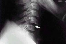
A cervical vertebral subluxation is defined when the bodies of two adjacent vertebrae are displaced relative to each other while still in contact, but the natural anatomical location of their articular surfaces is disrupted.
Epidemiology
According to some reports, traumatic cervical vertebral subluxations account for 45-60% of cases, with more than half of these injuries related to motor vehicle accidents and about 40% related to falls.
Adult cervical subluxation usually occurs in the lower cervical segments (C4-C7). Acceleration/deceleration trauma and direct impact to the neck cause subluxation at the level of the C4-C5 vertebrae in 28-30% of cases; half of anterior neck subluxations involve the C5-C6 vertebrae.
In young children - due to the anatomical characteristics of the developing spine - cervical vertebrae subluxation occurs in its upper cervical region (C1-C2) in about 55% of cases.
A very rare injury is a subluxation at the level of the C2-C3 vertebrae. [1]
Causes of the cervical vertebrae subluxation
As the main causes of subluxation (in Latin - subluxation) of the vertebrae of the neck (C1-C7) experts call trauma to the cervical spine, in particular, strong blows to this area of the spinal column, as well as sharp tilting or tilting of the head - extensor injuries of III-VII cervical vertebrae.
Often the etiology of subluxations of the neck vertebrae is associated with cervical spine instability, which is characterized by hypermobility of the cervical vertebrae - when the amplitude of their movements exceeds the normal range. This is due to weakness of the ligamentous structures fixing the vertebrae: the anterior and posterior longitudinal ligaments, the yellow ligament between the arches of neighboring vertebrae, the intercostal ligaments, as well as the fibrocartilaginous intervertebral discs and their fibrous rings.
Cervical vertebral subluxation in newborns usually affects the C1 vertebra (atlantus) and the atlantoaxial joint - the junction of the atlantus and C2 (axis) - and occurs with rotational birth trauma of the cervical spine.
It should be noted that head tilt to the front and back (nodding), as well as lateral tilts and rotation (rotation) occur in the paired atlanto-occipital joints of the craniovertebral zone (articulations of the condyles of the occipital bone with the upper articular fossa of the C1 vertebra) and in the medial atlantoaxial joint that unites the C1 and C2 vertebrae with its denticle (dens axis). Neck flexion and extension and its lateral inclinations occur in the middle and lower part of the cervical spine, i.e. In the subaxial spine, which includes vertebrae from C3 to C7.
There are different degrees of displacement of the body of one vertebra relative to the neighboring vertebra and the articular surfaces of the vertebrae of the given section. Depending on this, degrees of subluxation are determined: displacement of up to 25% is a Grade I subluxation; 25% to 50% is a Grade II subluxation; and 50% to two-thirds is a Grade III subluxation. [2]
Risk factors
In addition to the fact that the cervical spine is most susceptible to injury (due to the limited strength of the cervical vertebrae, the oblique position of their articular surfaces, and the relative weakness of the muscles that provide neck movement), vertebrologists include risk factors for cervical vertebral subluxation:
- Various congenital anomalies of the cervical spine, including vertebral arch dysplasia; occipital assimilation of the atlas (partial or complete fusion of the C1 vertebra with the occipital bone of the skull); splitting of the anterior and posterior arches of the atlas (in skeletal dysplasias, Down, Goldenhar and Conradi syndromes); Klippel-Feil syndrome (with fusion of the vertebrae of the neck); bony septum on the posterior arch of the atlanta (Kimmerly's anomaly); separation of a part of the C2 vertebral dentition from its body - os odontoideum, characteristic of mucopolysaccharidosis type IV (Morquio syndrome);
- Axis tooth fractures (C2 vertebral dentition);
- Cervical osteochondrosis;
- Cervical spondylosis;
- Rheumatoid and reactive arthritis; [3]
- Juvenile ankylosing spondylitis;
- Disc protrusion;
- Undifferentiated connective tissue dysplasia, which leads to disruption of the structure of intervertebral discs and instability of the spinal column;
- Hypermobility (increased mobility) of the cervical vertebrae in Marfan syndrome or ehlers-Danlos syndrome (with weakness of the ligaments between the skull and the C1 and C2 cervical vertebrae).
Pathogenesis
In subluxations of the vertebrae of the neck, the pathogenesis of displacement of their articular surfaces is due to the action of external shear force or the combined effect of flexion and forced extension (distraction), which exceed the capabilities of the ligamentous structures fixing the vertebrae.
This results in partial disruption of vertebral fusion in the form of localized spinal deformity with a sharp curvature (angular kyphosis), anterior rotation of the vertebra, anterior narrowing and posterior expansion of the disc space between adjacent vertebrae, displacement of the articular facets of the vertebrae relative to the adjacent underlying planes, expansion of the intercostal space, etc.
Thus there are different types or categories of subluxations in the cervical spine: static intersegmental, kinetic intersegmental, sectional, and paravertebral.
Static intersegmental subluxation includes changes in interosseous distance, flexion and rotation disorders, anterior displacement (anterolisthesis) or posterior displacement (retrolisthesis), and foraminal impingement or stenosis of the spinal foramen (foramen vertebrale) where the spinal nerves pass.
In kinetic intersegmental subluxation, there is either hypermobility of the vertebrae and their aberrant (opposite) motion, or displacement and immobility of the facet (arcuate) intervertebral joints.
If the subluxation is sectional, specialists observe anomalies of cervical spine movement and curvature and/or unilateral inclination of its part. In cases of paravertebral subluxations, pathologic changes in the ligaments are noted. [4]
For more on the anatomical features of the cervical vertebrae, see. - anatomical and biomechanical features of the spine
Symptoms of the cervical vertebrae subluxation
Since the uppermost vertebra of the cervical spine has no body and is connected to the adjacent vertebra by its arches (anterior and posterior) and the C2 dentate process, subluxation of the C1 cervical vertebra (atlanta) and subluxation of the C2 cervical vertebra (axis) are considered by specialists as atlantoaxial subluxation (C1-C2 subluxation). Such a subluxation - with restricted mobility of the cervical spine - can occur when the neck is abruptly flexed. But in addition to traumatic origin, when subluxation of the cervical vertebra in a child, in particular, C1 is due to dislocation or fracture of the vertebra C2, disruption of the articulation of the atlantoaxial joint in children can be due to relaxation of its transverse ligament - Grisel syndrome, which is observed after inflammation of the soft tissues of the neck (peritonsillar or pharyngeal abscess), as well as after otorhinolaryngologic surgeries.
The symptoms of such subluxation are manifested by intense neck pain (irradiating to the chest and back), headaches in the occipital region, dizziness, and rigidity of the occipital muscles. In most cases, there is persistent torticollis and abnormal head posture with chin turning in one direction and neck tilt in the opposite direction.
Subluxation of the C3 cervical vertebra limits flexion and extension of the neck and can affect jaw movement, as well as cause loss of diaphragm function (due to injury to the diaphragmatic nerve at the C3-4-5 level), requiring the use of ventilators to maintain breathing. If the cervical nerve plexus (plexus cervicalis) is compressed, paralysis of the arms, trunk and legs may occur, as well as bladder and bowel control problems.
Subluxation of the C4 cervical vertebrae is similar. And with subluxation of the C5 cervical vertebra, there is difficulty or weakness in breathing, problems with the vocal cords (hoarseness), neck pain, limited mobility of the wrists or hands.
If there is a subluxation of the C6 cervical vertebra, patients experience: pain when turning and bending the neck (including shoulder pain); stiffness of the neck muscles; numbness and tingling (paresthesia) of the upper extremities - in the fingers, hands, wrists or forearms; there may be difficulty breathing and impaired bladder and bowel function.
The first signs of subluxation of the last cervical vertebra (C7) may manifest as a burning sensation and numbness in the arms and shoulders with impaired mobility, pupil constriction and partial ptosis; other manifestations are the same as in C6 subluxation.
Rotational subluxation of the cervical vertebra with its rotation around the frontal axis is discussed in detail in the publication - rotational subluxations of the atlantus
If the articular processes of the vertebrae slip when the neck is flexed, but when the neck is flexed, they return to their normal position, a so-called habitual cervical vertebral subluxation is diagnosed. Read more in the article - habitual atlantoaxial subluxation
The instability of the cervical spine and its deformation are often complicated by chronic rheumatoid arthritis, in which some patients have a long-standing subluxation of the cervical vertebrae, in most cases - anterior atlantoaxial, causing severe pain in the neck and occipital region of the head. [5]
Complications and consequences
Complications and consequences of cervical vertebral subluxations include:
- Pinched nerve in the cervical spine, in particular the occipital nerve, and the development of occipital neuralgia - with aching, burning or throbbing pain on one or both sides of the head, pain in the eye sockets and increased sensitivity to light, pain behind the ears;
- Diaphragmatic nerve injury with unexplained dyspnea; orthopnea (dyspnea occurring in a horizontal position); insomnia and increased daytime sleepiness; morning headaches, fatigue, and recurrent pneumonia;
- Acute, subacute or chronic spinal cord compression with paresthesia, loss of sensation and spastic paresis of the hands, quadriplegia, quadriparesis and cruciate palsy (bilateral paralysis of the upper extremities with minimal or no involvement of the lower extremities);
- Occlusive damage to the vertebral artery, which manifests as vertebral artery syndrome;
- The development of scoliosis of the cervical spine.
Subluxation of the cervical vertebra in newborns can lead to narrowing of the spinal canal and compression of the spinal cord with neurological disorders, in particular, paresis or paralysis of the limbs or signs of cerebral ischemia in newborns - due to compression of the large vertebral arteries. [6]
Diagnostics of the cervical vertebrae subluxation
Anamnesis, examination of the patient, recording of the patient's complaints, and visualization of the vertebral joints allow the diagnosis of cervical vertebral subluxations.
Instrumental diagnostics is performed using x-ray of the cervical spine (with determination of spondylometric parameters); computer or magnetic resonance imaging, vertebral artery angiography, electromyography. For more details, see. - spine Examination Methods
An integral part of the diagnosis is the neurologic evaluation of the patient by identifying motor weakness, level of areflexia, and the presence of concomitant gorner syndrome.
Differential diagnosisincludes cervical vertebral fracture, dislocation and pseudo-dislocation associated with the absence of the vertebral body pedicle (a cylindrical protrusion of hard bone and its dorsal part), as well as other conditions with a similar clinical picture, For example, neuralgia with nerve root impingement (which may be accompanied by cervical osteochondrosis and osteoarthritis), tuberculous spondylitis, labyrinth angiovertebrogenic syndrome, and others. [7]
What do need to examine?
Who to contact?
Treatment of the cervical vertebrae subluxation
The main method of treatment is to correct the subluxation of the cervical vertebra by gradual traction (traction) with the help of orthopedic devices (Glisson loop and more modern devices Halo Skeletal Fixation for reliable external fixation and stabilization of the cervical spine).
They use traction according to the Richet-Güter method, Gardner-Well traction (using a spring-loaded tensioning device), Halo-Gravity Traction, after which an immobilizing cervical orthosis should be worn for a certain period of time.
There is also a Singhal traction bed with a tensioner handle and strain gauge to create additional traction while flexing the cervical spine.
The new AtlasPROfilax technology using a special vibrating device is used to reposition the C1 vertebra.
In some cases, surgical fusion of two vertebrae - spondylosis - may be necessary to stabilize the cervical spine. And if there is disc prolapse, the next step is an anterior access with discectomy and open repositioning with a Caspar distractor. [8]
Prevention
In many cases, prevention of cervical spine injury with subsequent vertebral subluxation can be prevented by following workplace safety rules, traffic rules and transportation of children in special child car seats.
And with instability of the cervical spine is recommended to wear fixation orthoses, undergo courses of therapeutic massage and physiotherapy, physical therapy to strengthen the muscles and ligamentous apparatus of the vertebral joints of the neck.
Forecast
In cervical vertebral subluxation, the prognosis depends on the complications associated with it and the success of treatment. A significant proportion of patients have neurologic complications that negatively affect their quality of life.
Can I enlist in the army if I have a cervical vertebra subluxation? It depends on its etiology and neurological status. If the subluxation is associated with instability of the cervical spine and has led to neurological complications, it is not eligible for military service.
List of authoritative books and studies related to the study of cervical vertebral subluxation
- "Cervical Spine Injuries: Epidemiology, Classification and Treatment" - by Jens R. Chapman, Edward C. Benzel (Year: 2015)
- "Cervical Spine Surgery Challenges: Diagnosis and Management" - by Ziya L. Gokaslan, Laurence D. Rhines (Year: 2008)
- "Cervical Spine II: Marseille 1988" - by Georges Gautheret-Dejean, Pierre Kehr, Philippe Mestdagh (Year: 1988)
- "Atlas of Orthopedic Surgical Procedures of the Dog and Cat" - by Ann L. Johnson, Dianne Dunning (Year: 2009)
- "Cervical Spondylosis and Other Disorders of the Cervical Spine" - by Mario Boni (Year: 2015)
- "Cervical Spinal Stenosis: The Old and the New" - by Felix E. Diehn (Year: 2015)
- "Cervical Spine Surgery: Challenges and Controversies" - by Edward C. Benzel, Michael P. Steinmetz (Year: 2004)
- "Manual of Spine Surgery" - by William S. Hallowell, Scott H. Kozin (Year: 2017)
- "Operative Techniques: Spine Surgery" - by John Rhee (Year: 2017)
- "Orthopaedic Surgery: Principles of Diagnosis and Treatment" - by Sam W. Wiesel (Year: 2014)
Literature
Kotelnikov, G. P. Traumatology / edited by Kotelnikov G. P.., Mironov S. P. - Moscow: GEOTAR-Media, 2018.

