Medical expert of the article

New publications
Vertebral articulations
Last reviewed: 04.07.2025

All iLive content is medically reviewed or fact checked to ensure as much factual accuracy as possible.
We have strict sourcing guidelines and only link to reputable media sites, academic research institutions and, whenever possible, medically peer reviewed studies. Note that the numbers in parentheses ([1], [2], etc.) are clickable links to these studies.
If you feel that any of our content is inaccurate, out-of-date, or otherwise questionable, please select it and press Ctrl + Enter.
The bodies of adjacent vertebrae are connected by intervertebral discs (disci intervertebrales), or intervertebral symphyses (symphysis intervertebrales), and the arches and processes are connected by ligaments.
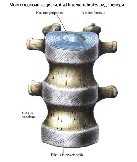
Each intervertebral disc is divided into a central and a peripheral part. The central part of the disc is called the nucleus pulposus, and the peripheral part is called the annulus fibrosus. The nucleus pulposus, which is a remnant of the spinal cord (chord), acts as a shock absorber between the bodies of two adjacent vertebrae. Sometimes there is a horizontal narrow gap inside the nucleus pulposus, which allows such a joint to be called a symphysis (half-joint). The peripheral part of the intervertebral disc (annulus fibrosus) is made of fibrous cartilage, which is firmly fused with the bodies of the vertebrae.
The thickness of the intervertebral disc depends on the level of its location and the mobility of the corresponding section of the spine. In the thoracic section, the least mobile, the thickness of the disc is 3-4 mm, in the cervical section, which has greater mobility, it is 5-6 mm; in the lumbar section, the thickness of the disc is 10-12 mm.
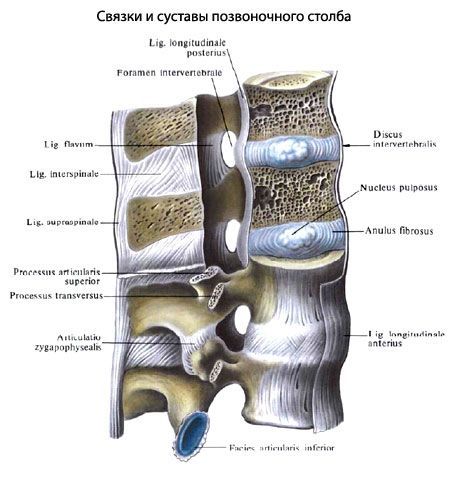
The connection of the vertebral bodies is reinforced by the anterior and posterior longitudinal ligaments.
The anterior longitudinal ligament (lig. longitudinale anterius) runs along the anterior surface of the vertebral bodies and intervertebral discs. This ligament begins on the pharyngeal tubercle of the occipital bone and the anterior tubercle of the anterior arch of the atlas and ends at the level of the 2nd-3rd transverse lines of the sacrum. The ligament firmly fuses with the intervertebral discs and loosely with the vertebral bodies.
The posterior longitudinal ligament (lig. longitudinale posterius) runs inside the spinal canal along the back surface of the vertebral bodies from the axial vertebra to the first coccygeal vertebra. At the level of the medial atlantoaxial joint, this ligament connects with the cruciate ligament of the atlas, and below it fuses with the intervertebral discs.
The arches of adjacent vertebrae are connected by yellow ligaments (ligg. flava), which consist mainly of elastic connective tissue that is yellowish in color. These ligaments are strong and elastic.

The articular processes form the zygapophysial (intervertebral) joints (artt zygapophysiales, s. intervertebrales). The planes of the joint spaces of these joints correspond to the orientation of the articular processes of the adjacent vertebrae of the cervical, thoracic and lumbar spine. The lumbosacral joints (artt. lumbosacrales), formed by the lower articular processes of the 5th lumbar vertebra and the upper articular processes of the sacrum, are considered separately.
All facet joints are flat, low-mobility joints, which is facilitated by the tight tension of the capsule, which is attached to the edges of the articular surfaces.
The spinous processes of the vertebrae are connected to each other by the interspinous ligaments (ligg. interspinale) and the supraspinous ligament (lig. supraspinale). The interspinous ligaments are thick fibrous plates located between the spinous processes. The supraspinous ligament is attached to the tops of the spinous processes of all vertebrae. In the cervical region, this ligament is called the nuchal ligament (lig. nuchae). The posterior edge of the supraspinous ligament is located between the external occipital protuberance above and the tops of the spinous processes of the vertebrae below.
Between the transverse processes of the vertebrae are located the intertransverse ligaments (ligg. intertransversaria) connecting them (Fig. 91). In the cervical spine, these ligaments are often absent.
The sacrococcygeal joint (art. sacrococcygea) is the connection of the apex of the sacrum with the 1st coccygeal vertebra. Often there is a gap in the intervertebral disc of this joint. The connection of the sacrum with the coccyx is strengthened by several ligaments. The paired lateral sacrococcygeal ligament (lig. sacrococcygeum laterale) goes from the lower edge of the lateral sacral crest to the transverse process of the 1st coccygeal vertebra. It is similar to the intertransverse ligaments. The ventral sacrococcygeal ligament (lig. sacrococcygeum ventrale) is a continuation of the anterior longitudinal ligament. The superficial dorsal sacrococcygeal ligament (lig. sacrococcygeum dorsale superficiale) goes from the edge of the sacral gap to the posterior surface of the coccyx. The deep dorsal sacrococcygeal ligament (lig. sacrococcygeum dorsale profundum), being a continuation of the posterior longitudinal ligament, is located on the posterior surface of the bodies of the 5th sacral and 1st coccygeal vertebrae. The sacral and coccygeal horns are connected to each other by connective tissue (syndesmoses). Mobility in the sacrococcygeal junction is more pronounced in women. During childbirth, some backward deviation of the coccyx is possible, which increases the size of the birth canal.
 [
[