Medical expert of the article

New publications
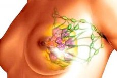
Postpartum mastitis
Last reviewed: 12.07.2025

All iLive content is medically reviewed or fact checked to ensure as much factual accuracy as possible.
We have strict sourcing guidelines and only link to reputable media sites, academic research institutions and, whenever possible, medically peer reviewed studies. Note that the numbers in parentheses ([1], [2], etc.) are clickable links to these studies.
If you feel that any of our content is inaccurate, out-of-date, or otherwise questionable, please select it and press Ctrl + Enter.
Lactational mastitis is defined as inflammation of breast tissue and commonly occurs in lactating women ( Amir et al., 2007 ). It is a painful condition with high fever; flu-like symptoms such as aches and chills; and red, tender, hot, and swollen areas of the breast ( Lawrence, 1989; World Health Organization, 2000 ). It is diagnosed symptomatically and there is no universally accepted clinical definition ( Zarshenas et al., 2017 ). Mastitis can present in a variety of patterns, from mild inflammation to more severe disease ( Michie et al., 2003 ).
Causes postpartum mastitis
There is no consensus regarding the etiology, which may be inflammatory, infectious, due to bacterial imbalance, or multifactorial (Baeza, 2016). Human milk is colonized by a wide variety of bacteria, some of which may originate from the maternal gut endogenously ( Marín, 2017 ). These commensal organisms appear to be important for the developing gut microbiome of infants. Potentially pathogenic bacteria have been isolated from breast milk of healthy lactating women, although there is evidence that some bacteria, particularly Staphylococcus aureus, are more common in women with mastitis than in women without it ( Hager et al. 1996; Kvist et al., 2008 ). Etiological theories include bacterial infection, such as through cracked nipples ( Foxman et al., 2002 ), or a dysbiotic process in which some species outgrow and others disappear ( Delgado, 2008 ). In addition, virulence factors, biofilm formation, antimicrobial resistance, and interactions with the host immune system are thought to play a role ( Contreras, 2011 ).
Pathogens
Symptoms postpartum mastitis
Patients complain of chills or rigors, weakness, headaches, sleep disturbances, appetite, pain in the mammary gland, and its enlargement. The clinical picture of the disease depends on the stage of the postpartum abscess.
- Pathological lactostasis develops on the 2nd-6th day after delivery. General health changes little. Body temperature rises to 38-38.5 °C. Uniform engorgement and pain in the mammary glands during palpation occur. Mastitis rarely develops without the lactostasis stage, but 8 to 30 days may pass between lactostasis and the first manifestations of serous mastitis, i.e. lactostasis is a latent stage of mastitis.
- Serous mastitis begins acutely. The general condition of the patient worsens. Headache, weakness, chills or rigors develop; body temperature rises to 38 °C. Gradually increasing pain in the mammary gland appears, especially during feeding. The skin in the affected area is slightly or moderately hyperemic. The mammary gland increases in volume; upon palpation, compacted areas of oval shape, dense elastic consistency, moderately painful are determined. The duration of this stage is 1-3 days. With inadequate treatment, serous mastitis becomes infiltrative.
- With infiltrative mastitis, the patient has a persistent fever, sleep and appetite are disturbed. More pronounced changes occur in the mammary gland: a dense, slightly mobile infiltrate is palpated under the altered area of the skin of the affected mammary gland, and regional axillary lymph nodes increase. The duration of this stage is 4–5 days, and if the infiltrate does not resolve, it becomes purulent.
- Purulent mastitis. The general condition of the patient is severe. Chills, an increase in body temperature to 39 °C and higher, complaints of poor sleep, loss of appetite are noted. The outlines of the affected mammary gland change depending on the localization and extent of the process, the skin of the gland is sharply hyperemic, its palpation is painful. The axillary lymph nodes enlarge and become painful upon palpation.
- The predominant form of purulent mastitis is infiltrative-purulent (in 60% of cases). The diffuse form is characterized by purulent impregnation of tissues without obvious abscess formation. In the nodular form, an isolated rounded infiltrate is formed without the formation of an abscess.
- Abscessing mastitis develops less frequently.
- Phlegmonous mastitis is an extensive diffuse purulent lesion of the mammary gland. It develops in every 6th-7th patient with purulent mastitis and is characterized by a very severe course. A sharp deterioration in the general condition, repeated chills, an increase in body temperature above 40 ° C are noted. Generalization of the infection with the transition to sepsis is possible.
- Gangrenous mastitis is an extremely rare and very severe form of the disease. Along with local manifestations, signs of severe intoxication are determined (dehydration, hyperthermia, tachycardia, tachypnea).
Currently, mastitis is characterized by a late onset, after the woman is discharged from the maternity hospital. Subclinical, latent forms of the disease are often detected, characterized by the lack of expression or absence of individual symptoms.
Stages
Postpartum mastitis is classified into stages.
- Pathological lactostasis (latent stage of mastitis).
- Serous mastitis.
- Infiltrative mastitis.
- Purulent mastitis.
- Infiltrative-purulent (diffuse, nodular).
- Abscessing (furunculosis of the areola, abscess of the areola, abscess in the thickness of the gland, retromammary abscess).
- Phlegmonous (purulent-necrotic).
- Gangrenous.
Complications and consequences
Most breast abscesses develop as a complication of lactational mastitis. The incidence of breast abscesses ranges from 0.4 to 11% among all lactating mothers. [ 11 ] Breast abscesses are more common in obese patients and smokers than in the general population. [ 12 ], [ 13 ]
Risk factors for the development of lactational abscess of the mammary gland include first pregnancy at a mother's age over 30 years, pregnancy of more than 41 weeks, and mastitis. [ 14 ] Breastfeeding women relatively often develop abscess of the mammary gland as a complication of mastitis. [ 15 ]
Mastitis can occur more than once, and women may experience lactational mastitis multiple times while breastfeeding the same infant. Women who develop mastitis may stop breastfeeding prematurely because of the pain the condition causes, fear that antibiotics may enter the milk, or inappropriate advice from health care providers to stop breastfeeding ( Foxman et al., 2002 ). This may expose infants to infection as well as increase the likelihood of obesity and metabolic disease later in life, particularly in low-income countries where there is a high disease burden and limited access to clean water and sanitation ( Dieterich et al., 2013 ). Thus, mastitis not only puts the mother at risk of more serious health complications, but may also result in potential loss of health benefits for the infant ( Wambach, 2003 ).
Diagnostics postpartum mastitis
- Complete blood count: leukocytosis, left shift in leukocyte count, increased erythrocyte sedimentation rate (ESR).
- Bacteriological examination of milk to determine the sensitivity of the pathogen to antibiotics. It is advisable to conduct the examination before the start of antibacterial therapy. Milk for examination is taken from the affected and healthy mammary glands. It is necessary to quantitatively determine the bacterial contamination of milk, since the diagnostic criterion for mastitis is the presence of 5x10 2 CFU/ml in milk.
- Ultrasound of the mammary glands: serous mastitis is characterized by a blurred tissue pattern, lactostasis; infiltrative mastitis - areas of homogeneous structure surrounded by an inflammation zone, lactostasis; purulent mastitis - dilated ducts and alveoli, with an infiltration zone around ("honeycomb"); abscessing mastitis - a cavity with uneven edges and bridges, surrounded by an infiltration zone.
Indications for consultation with other specialists
Consultation with a surgeon and anesthesiologist is indicated due to the need for surgical treatment of purulent and phlegmonous mastitis.
What do need to examine?
How to examine?
Who to contact?
Treatment postpartum mastitis
Lactational mastitis can be clinically characterized as "self-limiting" because it usually resolves without medical intervention through self-management, such as massaging the affected breast, feeding or expressing frequently enough to empty the affected breast, and using cold compresses to soothe the inflammation. ( Spencer, 2008; Wambach, 2003 ). However, some women require antibiotics to treat the infection, and if left untreated or untreated, infectious mastitis can lead to breast abscess or septicemia, which may require hospitalization and possibly surgery ( Thomsen et al., 1984 ).
Treatment goal:
- Eradication of the pathogen, relief of disease symptoms, normalization of laboratory parameters and functional disorders.
- Prevention of complications of the disease.
Indications for hospitalization
The appearance of clinical and laboratory signs of mastitis.
Non-drug treatment of postpartum mastitis
During illness, regardless of the clinical form, feeding the child from either the sick or healthy breast is unacceptable.
It is necessary to use a bandage that suspends the mammary gland and dry heat on the affected area. Physiotherapy
- In serous mastitis, microwaves of the decimeter or centimeter range, ultrasound, and UV rays are used; in infiltrative mastitis, the same physical factors are indicated, but with an increase in thermal load.
- In case of purulent mastitis after surgical treatment, first an UHF electric field is used in a low-thermal dose, then UV rays in suberythemal and low-erythemal doses.
Drug therapy
- Lactation should be slowed down or suppressed with the help of medications.
- In serous and infiltrative mastitis, lactation is inhibited, and if there is no effect from therapy within 2-3 days, it is suppressed. The consent of the mother must be obtained for lactation suppression.
- In case of purulent mastitis, lactation must always be suppressed.
- Depending on the severity of the clinical picture of the disease and the severity of lactation, cabergoline is used at a dose of 0.25 mg every 12 hours for 2 days or bromocriptine at 2.5 mg 2-3 times a day for a course of 2-14 days.
- Antibacterial therapy.
- The drugs of choice are penicillins (for example, oxacillin at a dose of 4 g/day intravenously, intramuscularly or orally).
- Cephalosporins of the first to third generations are effective.
- Cephalotin at a dose of 4–6 g/day intravenously or intramuscularly.
- Cefazolin at a dose of 4–6 g/day intravenously or intramuscularly.
- Cefuroxime at a dose of 4–6 g/day intravenously or intramuscularly.
- Cefotaxime at a dose of 4–6 g/day intravenously or intramuscularly.
- Cephalexin at a dose of 2 g/day intravenously or intramuscularly.
- In case of allergy to penicillins and cephalosporins, lincomycin is used at a dose of 1.8 g/day intravenously, intramuscularly.
- Aminoglycosides are effective: gentamicin at a dose of 0.12–0.24 g/day intramuscularly, amikacin at a dose of 0.9 g/day intravenously or intramuscularly, sisomicin at a dose of 3 mg/kg of body weight per day intravenously or intramuscularly, tobramycin at a dose of 3 mg/kg of body weight per day intravenously or intramuscularly.
- Medicines that increase specific immune reactivity and non-specific defense of the body.
- Antistaphylococcal human immunoglobulin, 100 IU every other day intramuscularly, in a course of 3–5 injections.
- Staphylococcal anatoxin, 1 ml at intervals of 3–4 days, 3 injections per course.
- Human normal immunoglobulin at a dose of 0.4–1 g/kg of body weight intravenously by drip daily for 1–4 days.
 [ 20 ], [ 21 ], [ 22 ], [ 23 ]
Surgical treatment of postpartum mastitis
In case of purulent mastitis, surgical treatment is indicated: it is necessary to perform a wide opening of the purulent focus with minimal trauma to the milk ducts. A radial incision is made from the border of the areola to the periphery. Bluntly destroy the bridges between the affected lobules, evacuate the pus, and remove necrotic tissue. Drainage is inserted into the wound. In case of phlegmonous and gangrenous mastitis, necrotic tissue is excised and removed.
Patient education
It is necessary to teach the mother how to properly care for her mammary glands, express milk, and feed the baby.
Further management of the patient
The question of resuming breastfeeding after mastitis should be decided individually, depending on the severity of the process and the results of bacteriological testing of breast milk.