Breast (breast)

The mammary gland (glandulae mammaris, s. Mamma, from the Greek mastos) is a paired organ, by origin is a modified sweat gland. In men, iron remains underdeveloped.
The mammary gland is located at the level of the III to IV rib, on the fascia that covers the large pectoral muscle, in connection with which it is also called the mammary gland. With the chest fascia, the mammary gland is loosely connected, which ensures its mobility. From the medial side, the mammary gland approaches the edge of the sternum with its base. Approximately in the middle of the gland is a nipple of the mammary gland (papilla mammaria) with pinholes on the apex, with which 10-15 excretory milk ducts (ductus lactiferi) open outward. A patch of skin around the nipple is a nipple (areola mammae), as well as a nipple, pigmented. In girls, it has a pink color, for women giving birth - brown (brown). The skin of the mug is uneven, on it are visible tubercles, on the surface of which the ducts of glands of the nipple gland (glandulae areolares) open, next to which the sebaceous glands are located. Bases of smooth muscle cells lie in the skin of the nipple and the nasal mug, part of which is oriented circularly, and a part - longitudinally. The contraction of these muscles strains the nipple.
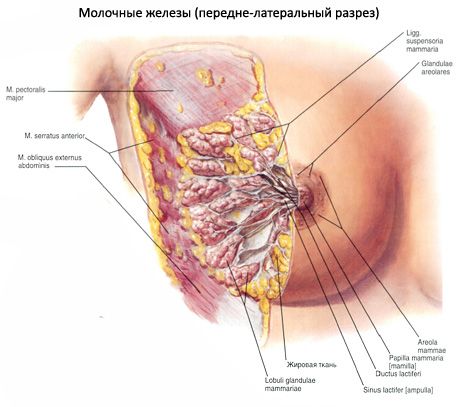
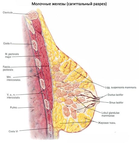
The body of the mammary gland (corpus mammae) consists of 15-20 lobes (lobi glandulae mammariae), separated from each other by interlayers of adipose tissue, permeated with bundles of loose fibrous connective tissue. These bundles pass into ligaments supporting the mammary gland (ligamenta suspensoria mammaria). The lobes consist of lobuli gl. Mammariae, having the structure of complex alveolar-tubular glands, which are arranged radially in relation to the nipple. The gland ducts (one from each lobe) open at the top of the nipple of the breast. On the way to the nipple (at its base) each duct has an extension - the lacteal sinus (sinus lactiferi).
In childhood, the mammary gland is underdeveloped, its maturation is confined to the period of puberty. When pregnancy glandular tissue grows, the gland increases in size. The nipple and the nipple circle darken. The dilated blood vessels (veins) appear through the thin skin of the gland. The maximum development of iron reaches the end of pregnancy. After lactation, the size of the gland is reduced. In the climacteric period, the iron undergoes partial involution. The function of the mammary gland is closely related to the activity of the gonads.
Anomalies in the development of the breast
There are cases of underdevelopment of one or both glands, there are additional (except for one pair) glands (polimastia - polymastia) or only additional nipples. In men, sometimes the glands develop according to the female type (ginaecomastia - gynecomastia).
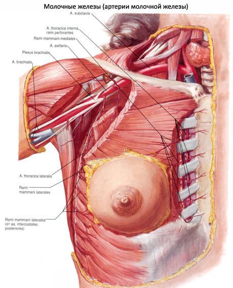
Vessels and nerves of the breast
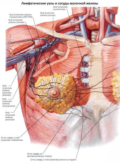
The branches of the 3-7th posterior intercostal arteries and the lateral thoracic branches of the internal thoracic artery are suitable for the mammary gland . Deep veins accompany the same-named arteries, the superficial veins are located under the skin, where they form a wide plexus plexus. Lymphatic vessels from the mammary gland are directed to the axillary nodes, the marginal nodes (on its and the opposite side) and the deep lower cervical (supraclavicular) lymph nodes. Sensitive innervation of the gland is carried out from intercostal nerves, supraclavicular nerves (from the cervical plexus). Along with the sensitive nerves and blood vessels, secretory (sympathetic) fibers enter the gland.
Last reviewed: 25.06.2018
Medical expert editor
Portnov Alexey Alexandrovich
Education: Kiev National Medical University. A.A. Bogomolets, Specialty - "General Medicine"