Medical expert of the article

New publications
Mammary gland (breast) examination
Last reviewed: 07.07.2025

All iLive content is medically reviewed or fact checked to ensure as much factual accuracy as possible.
We have strict sourcing guidelines and only link to reputable media sites, academic research institutions and, whenever possible, medically peer reviewed studies. Note that the numbers in parentheses ([1], [2], etc.) are clickable links to these studies.
If you feel that any of our content is inaccurate, out-of-date, or otherwise questionable, please select it and press Ctrl + Enter.
Examination and palpation of the mammary glands by a gynecologist is as necessary as examination of the cervix in a mirror during a gynecological examination.
When examining the mammary glands, it is necessary to pay attention to the structure of the mammary glands, their size (hypoplasia, hypertrophy, graphic changes).
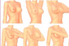
Examination of the mammary glands is performed in a standing and lying position with sequential palpation of the external and internal quadrants of the gland. Palpation allows one to determine the location of the tumor, its size, boundaries, consistency, and relationship with surrounding tissues. First, it is performed with light touches of the pads of the II, III, and IV fingers, placed flat on the palpated mammary gland.

Then they move on to deeper palpation, but it should also be painless. Examination of the marker gland in a horizontal position can significantly facilitate the diagnosis of minimal tumors. In this position, the entire mammary gland (breast) becomes softer, which allows identifying small areas of compaction in it. In addition, when the woman being examined is in a horizontal position, areas of dyshormonal hyperplasia become softer to the touch, or are not determined at all, while the tumor node does not change its consistency compared to the examination while standing.
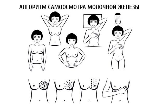
All patients are assessed for the presence or absence of discharge from the nipples, its color, and consistency. Palpation of the supraclavicular and subclavian areas, as well as the axillary areas, is performed to identify enlarged lymph nodes.
 [
[ What do need to examine?
How to examine?