Medical expert of the article

New publications
Breast exam
Last reviewed: 06.07.2025

All iLive content is medically reviewed or fact checked to ensure as much factual accuracy as possible.
We have strict sourcing guidelines and only link to reputable media sites, academic research institutions and, whenever possible, medically peer reviewed studies. Note that the numbers in parentheses ([1], [2], etc.) are clickable links to these studies.
If you feel that any of our content is inaccurate, out-of-date, or otherwise questionable, please select it and press Ctrl + Enter.
Examination and palpation of the mammary glands
Preventive examination of the mammary glands should be carried out monthly on the same day of the cycle, because in the mammary gland, as in the entire reproductive system, cyclical structural changes occur every month.
It is best to conduct a self-examination on the 5th-10th day of the cycle - it is during this period that the breasts are most relaxed and easily palpated. If the examination is carried out on a woman of climacteric age, it is recommended to do it on the same day of each month.
Self-examination is carried out in a room with sufficient lighting - most often this is done in a shower or bathroom. It is desirable to have a large mirror opposite. The examination usually does not take much time and over time turns into a familiar standard procedure.
Algorithm for examining the mammary gland
Part I – examining the linen.
- What can be seen on underwear, in particular, on a bra? These can be traces of small discharges from the glands, such as blood, pus, serous fluid, as well as already dried crusts. All these signs are of great importance in the diagnosis of breast diseases.
Part II – assessing the general condition of the mammary glands.
- We undress to the waist and stand directly in front of the mirror, arms down. We examine the left and right breasts for their size, asymmetry, and clarity of borders. After that, we evaluate the same thing, raising our arms up and holding them behind our head. We see if the shape of the breasts changes with body movements, if fluid is released from the nipples.
Part III – pay attention to the condition of the skin on the chest.
- We check the skin for elasticity, the ability to gather it into folds, the color and the presence of rashes and red spots. We examine the glands for the presence of diaper rash, areas like "lemon peel", ulcerated or wrinkled surfaces, retracted skin. The breasts are palpated alternately, in a vertical position, keeping the fingers of the hands closed.
Part IV – palpating the glands while standing in front of a mirror.
- This procedure is conveniently done standing in the shower and soaping your hands and chest. The left gland is palpated with the right hand, and vice versa. Palpation should be done with three or four closed fingers, first along the gland, then in a circle. If the breast is large, it is recommended to hold it with the free hand from below. To begin, palpate the surface of the breast, gradually going deeper into the tissue with the pads of the fingers. It is advisable to examine not only the glands themselves in this way, but also the area from the clavicular region to the lower rib, as well as from the middle of the chest to the armpit area. Enlarged lymph nodes are often found in this place.
Part V – palpate the chest while lying on your back.
- We lie down on a hard and flat surface, the floor is fine. One hand is behind the head, and the other one feels the opposite breast. It is recommended to conduct the examination with spiral movements, from the armpit area to the areola.
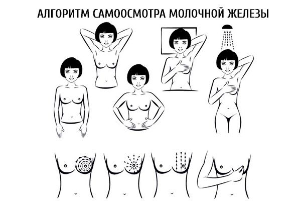
Part VI – we examine the nipple of each gland.
- When examining the nipples, you need to pay attention to their shape and shade, the presence of indentations, ulcers, erosions and cracks. It is advisable to feel not only the nipple itself, but also the area around it. Additionally, at the end of the examination, you should carefully grasp the nipple with two fingers and squeeze it slightly, checking for discharge.
After the self-examination, if you suddenly find any suspicious elements or signs, be sure to contact a doctor - a gynecologist or mammologist. Do not try to diagnose yourself and especially begin treatment. Only a qualified medical specialist should do this.
 [ 5 ], [ 6 ], [ 7 ], [ 8 ], [ 9 ], [ 10 ], [ 11 ]
Examination of the mammary glands in pregnant women
During pregnancy, it is also important to conduct a self-examination of the mammary glands. However, at this stage, it becomes more difficult to do this, because the breasts are growing, their sensitivity is increasing. Nevertheless, doctors advise conducting a self-examination at least once a month.
Before undergoing the examination, a pregnant woman should take into account all physiological changes in the breast so as not to mistake them for pathology:
- the glands increase in volume;
- sensitivity increases, pain may appear;
- the area around the nipple darkens, which is associated with increased secretion of skin pigment;
- visible blood vessels in the chest area may darken (caused by increased blood circulation in the glands);
- yellowish viscous discharge from the nipples (colostrum) may appear;
- the nipples protrude, increase in size, the areolas also increase in diameter;
- Small bumps appear around the nipples – this is the enlargement of the gland openings.
During pregnancy, it is especially important to pay attention to the choice of underwear. The bra should have good breast support, wide straps. It is undesirable to have bones and other details that can pull and squeeze the breast.
Examination of the mammary glands in case of lactostasis
During lactation, a woman may sometimes experience milk stagnation - lactostasis, or blockage of the milk duct. How can a woman determine the development of lactostasis by self-examination?
With lactostasis, the gland generally has a soft consistency, but at the same time, there are areas of lumps, density, and soreness. Externally, these areas may look reddened. Milk flows out of all the lobes without problems, but it may flow out of one lobe with difficulty, or not at all. In such a situation, we can safely say that there is a blockage. If milk stops flowing out of one of the glands at all, while it becomes dense, tense, and full, then lactostasis must be diagnosed. Milk expression is extremely difficult or ineffective.
You can try to do a light massage of the affected breast. The massage should not be aggressive and rough, but soft, gentle, careful: it is allowed to tap with fingertips, stroke. If lactation is not restored, then you will have to see a doctor, otherwise the stagnant state can turn into a complicated inflammatory process - mastitis.
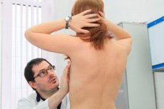
Examination of the mammary gland by a doctor
The doctor examines the patient's mammary glands more thoroughly, paying attention to many points that the woman herself often ignores (not because of inattention, but because of ignorance or inconvenience of performing some techniques).
The most frequently used technique by doctors is to change the position of the upper limbs of the woman being examined:
- First, the patient places her hands on top of her thighs (this helps to relax the chest muscles);
- then the patient presses her hands to her thighs (to tense the chest muscles);
- the woman is asked to raise her crossed arms above her head (definition of the umbilication symptom, which indicates the development of a cancerous tumor in the gland);
- The woman is asked to lean forward to relax the mammary glands (to assess the function of the suspensory ligaments).
Each breast area is examined carefully, along the entire length of the chest. At the same time, the doctor examines the armpit area, the inframammary area, and the surface of the anterior chest to the clavicular area. A typical examination includes palpation in certain areas, in circles, in a spiral, and also in radial directions from the nipple to the periphery. This approach allows not to miss a single area. The skin, subcutaneous fat, glandular tissue, and lymph nodes (in the axillary, supraclavicular, and subclavian areas) are palpated separately.
Examination of the mammary glands is a simple and accessible diagnostic method that should become a habit for every woman, especially after 35 years. Periodic examinations will help to detect the appearance of dangerous signs in time and begin treatment. Even oncological diseases are successfully cured if the course of therapy begins at an early stage of the pathology. If you have any suspicions, do not postpone visiting a doctor - this is the key to your healthy and fulfilling life.