Medical expert of the article
New publications
Pink blackheads
Last reviewed: 04.07.2025

All iLive content is medically reviewed or fact checked to ensure as much factual accuracy as possible.
We have strict sourcing guidelines and only link to reputable media sites, academic research institutions and, whenever possible, medically peer reviewed studies. Note that the numbers in parentheses ([1], [2], etc.) are clickable links to these studies.
If you feel that any of our content is inaccurate, out-of-date, or otherwise questionable, please select it and press Ctrl + Enter.

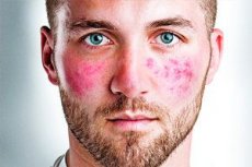
Rosacea (synonyms: acne rosacea, rosacea, red acne) is a chronic disease of the sebaceous glands and hair follicles of the facial skin in combination with increased sensitivity of the capillaries of the dermis to heat.
Causes pink acne
It is believed that rosacea is an angioneurosis in the innervation zone of the trigeminal nerve, caused by various factors: constitutional angiopathy, neurovegetative disorders, emotional stress, hormonal imbalance, dysfunction of the digestive tract, fecal infection.
Acne rosacea develops as a result of angiopathy and inflammatory reaction in the skin of the face under the provoking influence of a complex of various factors: endocrine disorders, liver diseases, gastrointestinal tract, vegetative-dystonia, alcohol abuse, etc. They occur mainly after 30 years. Acne glandularia can contribute to the development of the process, especially pustulosis, due to the cell-mediated immune response. Clinically manifested by stagnant erythema, telangiectasias and scattered papular-pustular rashes. In some cases, rashes can also be on other parts of the body (chest, back).
Some authors consider rhinophyma as one of the forms of rosacea, which is characterized by the development of lumpy, lobular nodules separated by grooves, sometimes reaching gigantic sizes, in the area of the nose, less often the chin and other areas. The following stages of the disease are distinguished: erythematous, papular, pustular and infiltrative-productive (rhinophyma). This division, however, is conditional, since patients usually have a combination of various morphological elements. Eye damage (blepharitis, conjunctivitis, iritis, keratitis) may be observed.
Rosacea-like changes in the skin of the face are observed in so-called perioral dermatitis, which is probably one of the forms of rosacea or seborrheides, developing mainly with prolonged use of fluorinated corticosteroid ointments.
In most patients, the presence of the mite "iron" is often found in the affected area.
Pathogenesis
In the erythematous-papular and papulopustular stages, focal lymphocytic infiltrates are observed in the dermis with the presence of reticular and mast cells, giant Lanhans cells, as well as hyperplasia of the sebaceous glands.
Pathomorphology
In the erythematous stage of the process, changes in the vascular apparatus of the skin predominate, then in the collagen substance. The vessels, especially the veins, are usually sharply dilated, loose fibrous connective tissue grows around their walls, without a pronounced inflammatory component, which indicates the presence of vasomotor disorders. Collagen fibers are loosened as a result of edema, hair follicles are somewhat atrophic with horny plugs in their mouths.
The papular stage is characterized by an inflammatory reaction in the form of a widespread or focal infiltrate of a lymphohistiocytic nature with the occasional presence of giant Pirogov-Langhans cells or foreign bodies.
In the pustular stage, changes in the vessels and follicular apparatus, a more intense inflammatory reaction are detected, expressed in massive infiltration by lymphocytes with an admixture of a large number of neutrophilic granulocytes, with the formation of pustules. Horny cysts, which are a consequence of atrophic changes in the follicular apparatus, as well as the destruction of collagen, are encountered more often than in the first two stages.
Rhinophyma is characterized by a pronounced proliferative component, characterized by the growth of connective tissue, leading to thickening of the dermis, obliteration of blood vessels, which further disrupts microcirculation in these areas. Sometimes inflammatory infiltrates with an admixture of neutrophilic granulocytes are detected.
 [ 13 ], [ 14 ], [ 15 ], [ 16 ]
Histogenesis
There are different points of view on the pathogenesis of acne rosacea. The most common opinion is about the important role of various neurotic disorders and vegetative dystonia, as well as stress influences. The role of hereditary predisposition is not excluded. There are works indicating the role of immune disorders. According to some authors, there is a deposition of IgM and/or complement in the dermal-epidermal junction and in dermal collagen. Circulating IgM antibodies were detected in the blood serum. Immunomorphological analysis of the infiltrate cells showed that the infiltrate consists mainly of LEU-1-reactive T cells with a predominant content of KEU-3a-antibody-positive T helper cells, while LEU-2a-cynecotic T cells were rare. These cells infiltrate the follicular epithelium and epidermis. In cases of the presence of demodex, the majority of T cells are found in infiltrates located around the mite and are T helper cells. The predominance of such T cells in the infiltrate in association with demodex indicates a violation of cellular immunity.
Symptoms pink acne
The disease begins with diffuse erythema of the face and telangiectasia. Against this background, in the presence of seborrheic phenomena, follicular nodules and scattered pustules appear. Papules and nodes have round and dome-shaped forms.
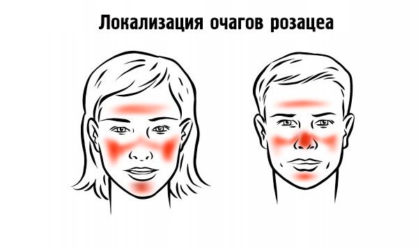
The elements are localized randomly on the skin of the nose, cheeks, chin, and less often on the neck, chest, back, and scalp.

Subjective sensations are insignificant: patients are concerned about the cosmetic defect and external resemblance to alcoholics. During a hot flash, redness of the face with a feeling of heat is noted. With a long-term course of the process and the absence of treatment, rhinophyma (pineal nose), metophyma (pillow-shaped thickening of the skin of the forehead), blepharophyma (thickening of the eyelids due to hyperplasia of the sebaceous glands), otophyma (growth of the earlobe in the form of a cauliflower), gnathophyma (thickening of the skin of the chin) occur.
Chronic blephoritis, conjunctivitis and episcleritis result in redness of the eyes. Keratitis and corneal ulcers are possible.
Stages
The following stages of the disease are distinguished:
- prodromal period - hot flashes;
- the first stage is the appearance of persistent erythema, telangiectasia;
- the second stage - the appearance of papules and small pustules against the background of persistent erythema and telangiectasia;
- the third stage - the appearance of a dense network of telangiectasia, papules, pustules against the background of persistent saturated erythema; there are nodes and extensive infiltrates.
 [ 17 ]
What do need to examine?
How to examine?
Who to contact?
Treatment pink acne
Complex treatment is carried out, including general and local medications. In case of abundant pustular rashes, antibiotics are prescribed (tetracycline 1-1.5 g/day in several doses, as the condition improves, the dose is gradually reduced to 250-500 mg once a day, or doxycycline 100 mg 2 times a day).
An important place is occupied by vitamin therapy (A, C, PP, group B) as a general tonic and for increasing capillary resistance. Trichopolum (metronidazole) has a good effect at 500 mg once a day during the first month, then 250 mg once a day during the next month. In case of a torpid course, immunomodulatory therapy is indicated. In case of a severe course of the disease and the absence of an effect from the above-mentioned agents, Roaccutane (isotretinoin) is indicated from 0.1 to 1 mg/kg of the patient's weight, depending on the clinical picture of the disease. In addition, depending on the degree of nervous system disorder, sedatives and tranquilizers are prescribed. It is also necessary to treat somatic pathology.
Topically, 0.75% cream or trichopolum gel are prescribed 2 times a day and antibiotics (clindomycin sulfate or erythromycin) in the form of a cream or ointment. If rosacea is accompanied by pronounced inflammatory phenomena, corticosteroid ointments are recommended. Considering that the mites "iron" support the inflammatory process, 20-30% sulfur ointment, the Demyanovich method, Skinoren cream, etc. are prescribed.
In sunny weather, sunscreen creams should be used.
More information of the treatment
Drugs

