Medical expert of the article

New publications
Physical methods of examining the patient
Last reviewed: 06.07.2025

All iLive content is medically reviewed or fact checked to ensure as much factual accuracy as possible.
We have strict sourcing guidelines and only link to reputable media sites, academic research institutions and, whenever possible, medically peer reviewed studies. Note that the numbers in parentheses ([1], [2], etc.) are clickable links to these studies.
If you feel that any of our content is inaccurate, out-of-date, or otherwise questionable, please select it and press Ctrl + Enter.
Physical research methods include those in which the doctor uses only his sense organs.
Questioning the patient provides significant information, which often allows for a diagnosis and treatment decisions. In other cases, questioning allows the doctor to make a preliminary conclusion and, when moving on to an objective examination, to focus on assessing the condition of certain organs whose damage seems most likely. There may also be situations when the patient is found unconscious and there is virtually no anamnesis data. In this case, a general examination may be ineffective and certain additional examination methods (for example, determining blood sugar levels) can be helpful.
Objective examination by physical methods most often provides essential information that has important diagnostic, prognostic and therapeutic value. As already mentioned, some symptoms can be detected only if they are thought about and specifically looked for. But there are often cases when only observation and examination of the patient in dynamics allow solving diagnostic and other problems, since a number of signs may appear at a later stage of the disease. In addition, it is necessary to take into account the possibility of the occurrence of some symptoms associated with the drug therapy.
Based on the results of a comprehensive study, it is possible to judge the general condition of the patient, which is characterized as satisfactory, moderate or severe. At the same time, sometimes the patient's well-being remains satisfactory or even good, despite the fact that his general condition can be assessed as moderate due to the presence of pronounced changes detected, for example, on an electrocardiogram (signs of acute infarction) or during a blood test (hyperkalemia).
The following are physical research methods:
- inspection;
- palpation;
- percussion;
- listening.
To indicate the position of organs or localization of changes detected during research using the above methods, it is advisable to focus on certain generally accepted points and lines, as well as on natural anatomical formations. Among the latter, the following should be noted:
- collarbones;
- costal arches and ribs;
- sternum, including manubrium, body, xiphoid process;
- spinous processes of the vertebrae, the counting of which is easy to begin with the clearly protruding 7th cervical vertebra;
- shoulder blades;
- iliac crests;
- pubic junction.
The following areas need to be kept in mind:
- jugular notch above the manubrium of the sternum;
- supra- and subclavian fossae;
- armpits;
- epigastric, or epigastric, region;
- subcostal regions, or hypochondria;
- lumbar region;
- groin areas.
In addition, the following vertical lines are used in physical examination:
- the anterior midline runs along the midline of the sternum;
- sternal, or sternal, lines run along the edges of the sternum;
- nipple, or midclavicular, lines;
- parasternal, or parasternal, lines are drawn in the middle between the two previous ones;
- the anterior axillary lines run along the anterior edge of the axillary fossa;
- the midaxillary lines pass through the center of the axillary fossa;
- the posterior axillary lines run through the posterior edge of the axillary fossa;
- the scapular lines pass through the lower angle of the scapula;
- the vertebral line runs along the spinous processes of the vertebrae;
- paravertebral lines.
A general examination is combined with a local examination (primarily of the skin), as well as palpation, tapping, and listening.
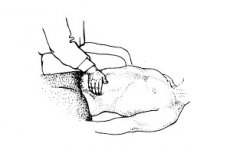
Palpation
Palpation of various organs and systems has its own characteristics, which are described in the sections devoted to the study of the corresponding systems. When palpating, the doctor always uses the information obtained during the previous examination of the patient and his knowledge of topographic anatomy. As A. L. Myasnikov wrote, it is always necessary to "call upon logical judgment, while palpating, think, and while thinking, palpate."
For effective palpation, it is necessary first of all for the patient to take a comfortable position, which is determined by the purpose of palpation. The position of the doctor should also be comfortable. It is advisable for the doctor to sit to the right of the patient's bed, facing him. The hands of the examiner should be warm, nails trimmed. The entire palmar surface of the hand is involved in palpation, although palpating movements are mainly performed with the fingers.
When palpating the abdominal cavity, it is important to use breathing movements.
Read also about palpation methods:
 [
[ Tapping (percussion)
The introduction of percussion into everyday medical practice was largely facilitated by J. Corvisart, a famous French physician and physician-in-ordinary to Napoleon I. Thanks to J. Corvisart, doctors became familiar with the work of the Viennese physician L. Auenbrugger, translated into French by him, “A New Method for Percussing the Human Chest to Detect Hidden Diseases Inside the Chest,” published in 1761.
When percussing the human body, different sounds are produced, the nature of which depends on the elasticity, air content and elastic tissue in the underlying organ.
A distinction is made between direct and indirect percussion, including the use of a special pleximeter - a plate and a hammer.
Currently, finger-on-finger percussion remains widespread, when the middle finger of the left hand is used as a pleximeter. It is firmly, but without pressure, applied to the percussed area. Tapping is performed with the middle finger of the right hand, which is slightly bent and does not touch the other fingers. The blow is applied to the middle phalanx of the pleximeter finger of the left hand, and the movement is performed mainly in the wrist joint (and not in the metacarpophalangeal joint) of the right hand. The force of the blow depends on the purpose and method of percussion. Louder percussion is also designated as deep, quiet - as superficial. While striking, the doctor listens to the sounds that arise, compares them and evaluates them, making a conclusion about the condition of the underlying organs, their boundaries.
Percussion can be comparative and topographic. Percussion is called comparative when the sounds obtained over anatomically identically located symmetrical areas of the body surface are compared (for example, percussion of the right and left lungs).
Topographic percussion aims to differentiate between different anatomical structures. The boundary between organs can be determined when they have differences in air content.
The following types of percussion sound are distinguished:
- loud - clear pulmonary;
- quiet - dull;
- tympanic.
A loud or clear percussion sound is normally obtained when tapping the chest above the lung area. It is determined by both the air content of the tissue and the content of a large number of elastic elements (alveolar tissue). A quiet or dull sound is normally obtained when percussing airless and soft organs that do not have elasticity, such as the heart, liver, and muscles. A distinction is made between percussion sounds of intermediate strength - dull or muffled (shortened).
In pathology, a clear sound becomes dull and dull due to a decrease or disappearance of air in the percussed organ.
The tympanic sound resembles the sound of a drum (tympanon) and is characterized by a higher pitch. It is obtained by percussion of air-containing smooth-walled cavities and over hollow organs containing air (stomach, intestines).
Thus, normally, a clear pulmonary sound is determined over the surface of the human body during percussion of the lungs, a dull quiet sound during percussion of the liver, heart and thick layer of muscles, and a tympanic sound over the abdominal cavity.
Read also about palpation methods:
Listening (auscultation)
Auscultation is the listening of sounds that occur naturally in the body, usually as a result of air or blood movement.
This method of research has been used for a very long time. The foundations of modern ideas about the importance of auscultation were developed by the great French doctor René Théophile Hyacinthe Laennec (1781 - 1826). He also suggested using a special device, a stethoscope, for this purpose. This idea came to R. Laennec in 1816. When examining a very overweight woman, he experienced difficulties in conducting direct auscultation. Taking a notebook and twisting it into a tube, he placed one end of this tube on the patient's heart area, and placed his ear on the other end. The quality of the sounds heard improved significantly.
The auscultation stethoscope was originally a wooden tube with different shaped extensions at both ends. Then came more comfortable soft stethoscopes that also amplify sounds.
A phonendoscope is a stethoscope whose end, which is placed on the patient's body, is covered with a membrane (usually made of plastic). This creates a small chamber that amplifies the sound.
Phonendoscopes and soft stethoscopes have slightly different designs and are made from different materials, although individual selection is possible.
When listening, it is important that the room is quiet. The stethoscope should be applied tightly enough. It should be borne in mind that sounds may occur due to the phonendoscope coming into contact with the hair on the body surface. In the case of significant hairiness, the areas corresponding to listening should be moistened to reduce additional sounds.
Auscultation is used in the study of the lungs and heart, where sound vibrations associated with their functioning naturally arise. Changes in the auscultatory picture, in particular the appearance of additional sounds, can be of decisive (key) importance in diagnosing the disease. It is important to know the normal variants. In addition, significant information can be obtained with dynamic auscultation and the appearance of new phenomena.
It should be borne in mind that auscultation is used after questioning and examining the patient, as well as palpation and percussion, which provide significant information for diagnosis and certain assumptions about the nature of the disease. Therefore, it is important that auscultation is performed purposefully, taking into account these assumptions.