Medical expert of the article

New publications
Examination and palpation of the heart
Last reviewed: 04.07.2025

All iLive content is medically reviewed or fact checked to ensure as much factual accuracy as possible.
We have strict sourcing guidelines and only link to reputable media sites, academic research institutions and, whenever possible, medically peer reviewed studies. Note that the numbers in parentheses ([1], [2], etc.) are clickable links to these studies.
If you feel that any of our content is inaccurate, out-of-date, or otherwise questionable, please select it and press Ctrl + Enter.
A general examination may be decisive for the diagnosis. The patient's sitting position or with the head of the bed elevated (orthopnea) is a characteristic symptom of heart failure with pulmonary congestion. In this case, the blood flow from the systemic circulation and the phenomena of congestion are reduced. Sometimes it is necessary to specifically ask the patient whether it is easier for him to breathe with the head of the bed elevated. In exudative pericarditis, patients sometimes sit leaning forward.
General inspection
Constitution (body build) is of relatively little importance for diagnosis, but stocky men (hypersthenics) are considered more likely candidates for coronary heart disease. Very tall, thin men with long fingers may have heart disease (aortic defect) at an early age, which is considered one of the signs of Marfan syndrome.
The skin and mucous membranes often change in heart disease. The most characteristic symptom is cyanosis - a bluish coloration of the skin, especially the fingers, tip of the nose, lips, ears - acrocyanosis. Cyanosis can be more widespread and significantly increases with physical exertion, which is accompanied by cold skin (in contrast to warm cyanosis in patients with pulmonary insufficiency). As with lung diseases, cardiac cyanosis is associated with a decrease in hemoglobin oxygenation, an increase in the circulation of reduced hemoglobin. In heart disease, there is a more active extraction of oxygen from oxyhemoglobin in peripheral tissues.
In long-term heart failure with liver congestion, jaundice may develop, which is combined with cyanosis. Petechial hemorrhagic rashes on the extremities, a peculiar skin color reminiscent of the color of coffee with milk, give reason to assume infective endocarditis, especially in patients with pre-existing valvular heart disease. Xanthelasma - slightly elevated, whitish spots on the skin of the eyelids - are associated with cholesterol deposition and lipid metabolism disorder, which is characteristic of coronary atherosclerosis. Some significance is attached to premature graying and baldness, which is often found among young patients suffering from ischemic heart disease.
Subcutaneous fat tissue and its expression have a certain significance. Its excessive development, general corpulence are an important risk factor for atherosclerosis. Exhaustion is observed in severe dystrophic stage of heart failure. Edema of the legs, especially shins and feet, is a characteristic sign of stagnation in the systemic circulation. Edema of one of the shins is characteristic of phlebitis of the deep veins of the shins. To detect it, it is useful to measure the circumference of the shins at the same level, while on the side of phlebitis the circumference will be larger.
Examination of the extremities sometimes provides significant data. Clubbing of the fingers and toes occurs in cyanotic congenital heart defects, as well as in infective endocarditis. Characteristic external changes in the skin and various joints can be detected in many diseases (e.g., systemic lupus, scleroderma, thyrotoxicosis, etc.), often accompanied by heart disease.
Changes in the lungs during heart failure are expressed in increased respiratory rate and the appearance of moist, silent wheezing in the lower lateral and posterior sections.
Examination of the heart area
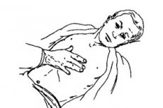
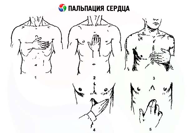
It is better to perform simultaneously with palpation, which, in particular, facilitates the detection of pulsations. Some pulsations are better perceived visually, others mainly by palpation. During examination, a cardiac hump can be detected, associated with deformation of the chest as a result of early enlargement of the chambers of the heart due to its defect. The most important pulsations in the heart area are the apical impulse and cardiac impulse, which can be used to judge hypertrophy and enlargement of the left and right ventricles of the heart, respectively.
The apical impulse is visible in most healthy people in the fifth intercostal space, 1 cm inward from the midclavicular line. To determine it, the palm of the right hand is placed on the specified area, and then the characteristics of the apical impulse are clarified using the fingertips of the right hand, with which its width, height, and resistance are established. Usually it is determined on an area of 1-2 cm 2. The apical impulse is associated not only with the contraction of the left ventricle, but to a greater extent with the rotation of the heart around its axis, which leads to a jerky movement of the heart towards the chest. The apical impulse is not visible and palpable if its localization corresponds to the rib (and not the intercostal space), as well as with severe pulmonary emphysema. An increase in the size of the apical impulse of more than 3 cm in diameter corresponds to dilation of the left ventricle. Strengthening (increased amplitude) and increasing resistance of the apical impulse correspond to left ventricular hypertrophy. In both cases, a shift of the apical impulse outward from the midclavicular line is simultaneously noted, and with pronounced hypertrophy and dilation even in the sixth intercostal space.
The cardiac impulse is determined outward from the left edge of the sternum at the level of the fourth rib and the fourth intercostal space. Normally, it is usually not visible and is not palpated or is determined with great difficulty in thin individuals with wide intercostal spaces. It begins to be clearly detected with hypertrophy of the right ventricle, with the systole of which it is associated. With severe pulmonary emphysema, the cardiac impulse may be absent even with significant hypertrophy of the right ventricle. In this case, pulsation in the epigastric region may be determined, which may be associated with pulsation of the aorta or liver.
A widespread cardiac pulsation can be determined slightly inward from the apical impulse in patients with transmural infarction, with aneurysm of the left ventricle.
Tremors of the chest wall in a limited area corresponding to the listening point of one or another valve can be detected in case of heart defects. This tremor is called "cat's purr" because it resembles the sensation that occurs when stroking a purring cat. This symptom practically corresponds to the vibrations that cause the appearance of a murmur in the heart due to the difficulty of blood flow through the atrioventricular and aortic openings during systole or diastole. Accordingly, the tremor can be systolic or diastolic. At the same time, the corresponding noise characteristic of the defect is heard. For example, diastolic tremor at the apex of the heart is determined in mitral stenosis simultaneously with diastolic murmur.
When the pressure in the large vessels (aorta or pulmonary artery) increases, the corresponding semilunar valves close more quickly at the beginning of diastole. This causes a small palpable push at the edge of the sternum in the first - second intercostal spaces, respectively on the left due to the closure of the pulmonary artery valves and on the right as a result of the slamming of the aortic valves.
Pulsation in the second intercostal space to the right of the sternum or behind the manubrium of the sternum may be determined during the development of an aneurysm of the aortic arch. Pulsation of the abdominal aorta may be detected in thin patients in the epigastric region and below.
Currently, precordial pulsation at various points can be recorded using special equipment in the form of a curve (kinetocardiogram), the analysis of which allows us to establish disturbances in the movement of the heart wall in different phases of the cardiac cycle.