Medical expert of the article

New publications
Right ventricular hypertrophy
Last reviewed: 05.07.2025

All iLive content is medically reviewed or fact checked to ensure as much factual accuracy as possible.
We have strict sourcing guidelines and only link to reputable media sites, academic research institutions and, whenever possible, medically peer reviewed studies. Note that the numbers in parentheses ([1], [2], etc.) are clickable links to these studies.
If you feel that any of our content is inaccurate, out-of-date, or otherwise questionable, please select it and press Ctrl + Enter.
The heart. How many beats does it make during a person's life, how much life-giving fluid, in the literal sense of the word, does it pump. But time comes and, like any mechanism, it also begins to give in. Right ventricular hypertrophy (right ventricular myocardial hypertrophy) is a condition when the size of the right ventricle of the heart increases, muscle tissue builds up, thereby increasing the load on the human blood pump itself - the heart.
As everyone knows from a school anatomy course, the human heart consists of four chambers. Two right valves with chambers are responsible in the human body for the normal functioning of the so-called small pulmonary circulation. The remaining left chambers pump blood plasma through the large systemic circulation. Therefore, in a healthy person, the so-called pulmonary pressure is lower than the venous pressure. When measuring arterial pressure, it is characterized by the lower figure in the readings. This disease is manifested by the fact that the lower figure of the tonometer readings increases, the difference in the pressure drop of the large and small circles shown by this device decreases, which contributes to the deterioration of the general condition of the person and, subsequently, to the persistent formation of the disease.
 [
[ Causes of Right Ventricular Hypertrophy
This disease cannot be called typical. It is not common, and sometimes it is difficult to recognize it. What are the causes of this disease?
There are two main causes of right ventricular hypertrophy. These are:
- Mitral stenosis, which is characterized by a decrease in the area of the opening that connects the right atrium and the ventricle of the same name. This opening closes the mitral valve.
- A heart pathology that formed in the womb.
That is, hypertrophy of the right ventricle develops on the basis of all sorts of deviations in the structure of the heart, often acquired at the stage of fetal development - this is in children, and in adults, the basis for the development of the disease can be any lung disease with complications that affect the heart muscle, or valvular heart disease.
Depending on the degree of progression of the disease and the characteristics of its genesis, cardiologists classify right ventricular hypertrophy into several types:
- Tetralogy of Fallot. This pathology manifests itself already with the birth of the child. Its symptoms can accompany the baby throughout the first year of life. Manifestations of this disease are also called "blue baby syndrome" - which is a manifestation of dysfunction of the blood outflow.
- Hypertension of pulmonary genesis. It is caused by an increase in pressure in the small pulmonary artery circle. In this connection, the patient develops shortness of breath, dizziness in combination with fainting states.
- Stenosis of the valve of the small ring of circulation. The manifestation of this pathology is a violation in the outflow of blood plasma into the blood vessel from the valve.
- Pathology of the interventricular septum. Defective structure of the cardiac septum allows two flows of adjacent sections to mix. This causes a decrease in the amount of oxygen carried, as well as an increase in the load on all areas of the heart, including the right ventricle.
Among the pulmonary pathologies that can cause right ventricular hypertrophy, the following can be particularly distinguished:
- Inflammation of the lungs or pneumonia.
- Fibrosis. On the contrary, the compaction of lung tissue formed as a result of an inflammatory process or for any other reason.
- Bronchial asthma.
- Emphysema is a pathological enlargement of the alveoli (lung sacs) and the airways adjacent to them.
- Chronic bronchitis.
- Pneumosclerosis. The growth of lung tissue, which may be a consequence of the same inflammatory process.
Signs of right ventricular hypertrophy
Quite a large number of diseases have similar symptoms. And only a specialist (a therapist, and in many cases only a specialist) is able to correctly analyze them and make a diagnosis. Only a cardiologist can diagnose the deviation from the norm in question.
Such a disease as right ventricular hypertrophy can be attributed to quite rare pathologies. Therefore, even if you have an electrocardiogram, it is quite difficult to detect it, since the weight of the right ventricle in percentage terms is less (it is approximately a third of the weight of the left one), which allows the left, large, contour to prevail in the cardiogram readings.
Therefore, signs of right ventricular hypertrophy on the cardiogram are easily read only with a significant increase in the mass component of the right ventricle.
Based on the above, medicine distinguishes the following types of right ventricular hypertrophy:
- The case when the mass of the right region is significantly greater than the weight of the left ventricle is an acute hypertrophy.
- Moderate pathology. Against the background of increasing parameters of the right heart, excitatory processes begin to proceed more slowly.
- Mild degree of the disease. Pathology of the right compartment is insignificant.
- In the early stages of the disease (right ventricular hypertrophy), the symptoms are weakly expressed, their manifestations are blurred. But as the pathology develops and the size gradually increases, the symptoms become stable and recognizable:
- Sudden dizziness, even to the point of fainting.
- Shortness of breath, making it difficult to breathe. Such attacks are usually accompanied by pain in the chest area.
- Severe attacks of arrhythmia. Rapid heartbeat.
Clearly visible swelling of the lower extremities.
Right ventricular hypertrophy in a child
The growth of the heart muscle increases the load on the right side of the baby's heart, which is much worse and more serious than with the same pathology of the left side. The whole point is that the pulmonary pulmonary circulation, and, accordingly, the sections that serve it, are adapted for normal operation in the area of low pressures. If there is a discharge of blood fluid in larger than normal volumes by the left half of the heart or in the case of pulmonary artery stenosis, the pressure of the pulmonary circulation increases, and the load on the right side of the heart muscle automatically increases. And in order to cope with the increased load, the heart muscle of the right ventricle has no choice but to build up mass, increasing in size. In this case, hypertrophy of the right ventricle develops in a child.
Monitoring the maximum number of cases of the disease led doctors to the conclusion that this disease is much more common in children than in adults. In a small person, this disease can occur in the first days of his life and have a purely physiological nature, since during this period the load on this half of the heart increases significantly. But these cases are quite rare. The largest percentage of right ventricular hypertrophy still falls on cases of congenital heart disease, the symptoms of which appear in the first days of a child's life.
But not only the components of the heart are subject to increased stress, but also the vessels with arteries that are part of the pulmonary system. And if the increased stress persists for a sufficiently long time, then the vessels become harder, which triggers the procedure of vascular sclerosis. Which, in turn, leads to a decrease in the plasma patency of the pulmonary ring, the pressure in the pulmonary circle increases, leading to a disease that is called Eisenmenger's syndrome in medicine. And the symptoms of this disease are already irreversible. Drawing a conclusion from all of the above, it is necessary to understand that right ventricular hypertrophy is serious and the problem cannot be left to chance. In this situation, urgent medical intervention is necessary to prevent further unfavorable developments.
Therefore, if your child has been diagnosed with signs of this disease, do not despair or panic. Just contact a cardiologist and have your child undergo a full medical examination.
Right ventricular hypertrophy in a newborn
Various age categories are subject to an increase in the volume and mass characteristics of the ventricle, but, nevertheless, hypertrophy of the right ventricle in a newborn (the so-called congenital pathology - heart defect) in percentage terms occurs more often than all other cases.
Cardiologists believe that the cause of this disease in very young children, newborns, and babies is:
- increased stress that affects the right side of the heart while still in the womb or in the first days after birth.
- disruption of the function of blood outflow from the right ventricle, which leads to a congenital pathology - hypertrophy of the right ventricle.
- Anatomical defects of the cardiac septum can also lead to pathological changes in the blood supply system. That is, there is no hermetic separation of one cavity of the heart from another, which leads to mixing of blood flows. In this case, the blood is poorly saturated with oxygen, and, consequently, the entire human body as a whole does not receive enough of it, which leads to systemic pathology. And in order to compensate for the lack of oxygen in the organs, the heart has to work with greater effort. And the result is hypertrophy.
- Also, the cause of this pathology in newborns can be called pulmonary valve stenosis.
Young mothers should understand that if any symptoms deviate from the norm, they should not despair and diagnose themselves. It is better to contact your pediatrician as soon as possible, and he, if necessary, will refer you to a pediatric cardiologist, and only he can confirm or refute this diagnosis. The sooner you contact the clinic with your baby, the faster and more gentle methods your child will be treated.
Hypertrophy of the right and left ventricles
Hypertrophy of the right and left ventricles is, in some sense, a precursor to a more severe disease caused by an increase in the myocardium. At the same time, it is a complex pathology caused by a significant growth of cardiac muscle tissue, while the volumes of the ventricular cavities remain unchanged.
Left myocardial hypertrophy. The work of the left ventricle ensures the functionality of the large circle of blood circulation. If its work is disrupted, a person begins to feel:
- A pressing pain in the chest.
- Sudden onset of dizziness.
- Frequently recurring fainting spells.
- The patient feels a loss of strength and apathy.
- Sleep may be disrupted.
- Disturbances in the functioning of the human nervous system are observed.
- Arrhythmia appears.
- Shortness of breath creates breathing difficulties. Moreover, it occurs not only during physical exertion, but also at rest.
Right myocardial hypertrophy. Its consequences are more destructive for the patient's body, since the work of the right ventricle is responsible for the small circulation cycle, which has a normal working pressure lower than in the large circuit. Therefore, when the pressure in it increases, the body suffers much more. Through blood vessels, the small circulation cycle connects the work of the heart (its right ventricle) with the lungs, therefore, any problems that arise with the lungs immediately affect the heart muscle, leading to hypertrophy of the right ventricle.
Diagnosis of right ventricular hypertrophy
The diagnosis of any disease should be made by a doctor after conducting a full range of studies. Diagnosis of right ventricular hypertrophy includes:
- Physical examination - a doctor's examination. Often it is this that prompts the idea of a disease. A competent cardiologist is able to hear heart murmurs and disruptions in the working rhythm.
- Electrocardiography. But with the help of a cardiogram you can see only the rhythm disturbance, but not the size disturbance. That is, it is an indirect diagnosis.
- Analysis of patient complaints.
- Echocardiography. This method, using ultrasound, makes it possible to determine the parameters of the heart muscle, measure its thickness, identify a violation of the outflow of blood through defects, and assess their size. It makes it possible to measure the pressure in the ventricle. A fairly accurate method of determination.
- ECG.
- Cardiovisor. This device allows you to observe the heart's work in dynamics. It can be used at home.
- Identification of hereditary predisposition to disease.
- The risk group also includes people who are overweight, or, conversely, athletes who receive heavy loads during training and competitions, as well as owners of bad habits. They need to periodically undergo examination by a cardiologist for preventive purposes.
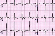
Right ventricular hypertrophy on ECG
Of course, only specialists with a medical education can and should read and decipher an electrocardiogram, but those who are particularly inquisitive, in order to broaden their horizons, can try to understand the physiological, impulse processes that occur in the myocardium with right ventricular hypertrophy.
Therefore, let's try to figure out what change right ventricular hypertrophy shows on the ECG. First, it is worth understanding that the mass component of the right ventricle is three times less than the mass of the left one, and in a normal state, the electrical impulses emitted by the half of interest to us are significantly lower. Therefore, in a healthy state, the signal from the left, "stronger" ventricle prevails. In a state of hypertrophy, the right ventricle begins to produce a stronger EMF, which shifts the total vector to the right.
In light of these findings, cardiologists distinguish between three types of right ventricular hypertrophy:
- Severe degree of hypertrophy. This type is characterized by the fact that the size of the right ventricle begins to exceed the corresponding parameters of the left.
- Medium degree of hypertrophy. In this case, hypertrophy of the right region already exists, but the parameters are still smaller than the dimensions of the left ventricle. The EMF of the right ventricle increases, but it is still weaker than the impulses that come from the left.
- Mild hypertrophy. There is a deviation from the norm of the right ventricle, but it is still insignificant.
Let's try to understand the symbols that appear on a cardiogram:
- The symbol P denotes the teeth responsible for the contraction of the atria.
- The letters Q, R, and S represent the characteristics of ventricular contraction.
- T is a characteristic of the relaxing signal in the ventricles of the heart.
Now let's figure out in what cases a cardiologist can make a diagnosis of right ventricular hypertrophy.
- If the electrocardiogram shows sufficiently high RV1,V2 teeth, while no deviations from the norm are observed in the bursts of the TV1,V2 teeth and the STV1,V2 segment.
- If, when the heart is working under load, the electrocardiogram displays high RV1, V2 bursts, while the pulsation of the STV1, V2 segment is reduced, and the amplitude of the T tooth V1, V2 has a negative value.
- The cardiologist states the presence of right ventricular hypertrophy with obvious signs of myocardial pathology and its increased overload if the combination of a high R crest with a reduced ST segment, as well as negative T values are seen not only in the V1, V2 areas, but also in other areas of the heart.
But it is worth noting that the ECG does not show right ventricular hypertrophy as clearly as left ventricular hypertrophy. Therefore, before making a final diagnosis, it is necessary to turn to other diagnostic methods.
What do need to examine?
Who to contact?
Treatment of right ventricular hypertrophy
Most often, hypertrophy of the right ventricle affects patients who have a history of chronic lung diseases, as well as heart defects acquired in the womb, etc. Treatment begins after diagnosis and establishment of the origins of the pathology.
Depending on the causes of changes in the myocardium of the right ventricle, the method used to treat right ventricular hypertrophy is used.
- The etiotropic method of treatment is used in case of a detected congenital heart defect. This method is aimed at eliminating or weakening the effect of the cause itself, which activates the disease.
- The pathogenetic method is used for "acquired" right ventricular hypertrophy. The method works to increase passive and active immune processes that block the cause of the disease.
Both of these methods work to normalize blood pressure, slow down the progression of the disease and, if possible, correct the defect. And the treatment should also be aimed at eliminating the immediate cause of the disease: be it chronic pulmonary diseases, congenital heart disease or pulmonary stenosis.
If right ventricular hypertrophy causes a heart defect, the patient will be shown surgical intervention. As a rule, this concerns small children. In this case, they try to perform the operation in the first year of the child's life.
If the cause of this disease is pulmonary, the doctor prescribes bronchodilators, respiratory analeptics, and mucolytics to the patient. Such as:
Bronholitin (bronchodilator). This drug is prescribed to an adult patient at the rate of one tablespoon three to four times a day.
For children over ten years old, the dosage is slightly smaller and amounts to a tablespoon (or two teaspoons) three times a day.
For children from three to ten years old, a single dose will be one teaspoon three times a day.
This medicine is not recommended for people suffering from angina, insomnia, glaucoma, heart failure, thyrotoxicosis and some other diseases. Broncholitin should not be given to children under three years of age, as well as to expectant mothers in the first trimester of pregnancy and during breastfeeding.
Analeptic (respiratory analeptics). Such drugs are used relatively rarely, but in case of asphyxia of a newborn, this medicine helps the baby to restore normal breathing. The baby is placed in a warm (38–39° C) bath. The mucus is sucked out of the baby’s nose with a special balloon. A solution is prepared from 1 ml of the drug and 5 ml of saline (it can be replaced with 5% glucose). The analeptic is given to the baby through a vein very slowly. If there is no obvious result, the medicine is administered again.
An analeptic should not be administered to a patient with epilepsy, convulsions, or tetanus.
Bromhexine (mucal agents). This drug is not given to children under six years of age in tablets. Children aged six to ten years are given 8 mg of the drug three times a day. When taking bromhexine, it is necessary to drink a large amount of liquid.
Toddlers under two years of age are given the drug in the form of syrup, 0.5 teaspoon. For children from two to six years of age, it is better to give the drug 0.5 - 1 teaspoon in the form of syrup. For older children (from six to 14 years) - 1 - 2 teaspoons.
To correct the blood pressure of a patient with right ventricular hypertrophy, a cardiologist may prescribe:
Euphyllin. The dosage is determined by the doctor individually. Adults are given 0.15 g at a time. There can be one to three such doses per day.
For children, the intake is spread over three approaches. The daily dosage is 7-10 mg per kilogram of the child's weight. If there is no obvious effect, the dosage can be increased until a positive result is obtained. But the dosage must be increased gradually, step by step, every two to three days. The course of treatment is determined by the doctor, and it can last from a couple of days to several months.
Euphyllin is contraindicated for people who are hypersensitive to the components of the drug, in case of arrhythmia, if the patient has a history of ulcerative diseases, heart failure, myocardial infarction and many others.
In the mild stage of right ventricular hypertrophy, the cardiologist may prescribe:
Nifedipine. The drug is taken two to three times a day at a dosage of 0.01 g. The dosage can be increased to 0.02 g. In very rare cases, the patient can receive 20 mg of the drug (0.02 g) four times a day, but the total daily dosage should not exceed 80 mg. The duration of the course is individual and is prescribed by the doctor based on the clinical picture of the disease and the patient's condition.
This medicine should not be taken in case of hypotension, acute heart failure, collapse, during pregnancy and lactation, as well as in case of some other diseases.
If the disease is in a decompensated mode, the patient receives medications of the nitrate group, such as nitrosorbide or nitroglycerin. The medications are taken under blood gas monitoring.
Nitrosorbide. An adult patient is prescribed a dosage of 5 to 10 mg of the drug half an hour before meals, three to four times a day. In severe cases of the disease, the dosage can be increased to 20 to 30 mg. If the patient has severe heart failure, he or she must take 20 mg (two tablets) every four to five hours.
This medicine is not prescribed for strokes, traumatic brain injuries, individual intolerance to nitrates, glaucoma and increased eye pressure, etc.
Nitroglycerin. If the drug is taken in tablet form, it is placed under the tongue until completely dissolved. It is used to quickly relieve acute pain. Nitroglycerin is prescribed in a dosage of one to two tablets (0.5 - 1 mg). But the total daily amount of the drug should not exceed 6 tablets.
Nitroglycerin in capsule form is consumed in the same way. To speed up the expected result, the capsule must be broken in the mouth with teeth. The amount of the drug taken depends on the frequency of pain attacks. Relief from angina symptoms and relief usually comes quickly, ½ to 2 minutes after taking the medicine. If there is no effect, then after five minutes you need to take another tablet. In the absence of a therapeutic result and after two or three tablets, you need to call an ambulance.
The list of contraindications is quite large. These include: individual intolerance to nitrates, recent head injury, acute myocardial infarction, toxic pulmonary edema, arterial hypotension, cerebral circulatory pathology, collapse and many other diseases. The full list of contraindications can be viewed in the instructions attached to the drug. Doctors do not prescribe nitroglycerin to children and adolescents under 18 years of age, as well as to mothers during pregnancy or lactation.
During the entire treatment period, the doctor must monitor the heart. And the patient will have to completely give up smoking and drinking alcohol during this time. At the same time, the patient must follow the daily routine and diet. Physical therapy and swimming will be useful.
Prevention of right ventricular hypertrophy
Every sensible person should understand that in order not to get a disease in any of its manifestations, it is necessary, first of all, to prevent or eliminate the cause of its occurrence. So, the prevention of right ventricular hypertrophy comes down to:
Carrying out activities that help prevent the progression of leg phlebothrombosis:
- Diagnosis of this pathology at early stages and immediate treatment.
- This also includes a preventive examination of hospital patients for increased risk of right ventricular hypertrophy.
- A postoperative patient diagnosed with leg phlebothrombosis should actively move (the blood should not "stagnate"), tie the operated leg with an elastic bandage. Follow the entire treatment protocol prescribed by the attending physician.
For chronic lung diseases:
- It is necessary to protect yourself from hypothermia and drafts.
- Quit smoking, avoid even passive smoke exposure.
- Don't let the disease get worse, but try to take action at the early stages of its manifestation.
- Lead an active lifestyle with moderate exercise.
- Oxygen cocktails can also be a good preventative measure.
Prognosis of right ventricular hypertrophy
Until recently, chronic pulmonary heart disease was considered an irreversible disease. Modern medicine classifies it as a reversible complication. Therefore, the prognosis for right ventricular hypertrophy today largely depends on the patient's medical history, the nature and severity of the disease that caused and triggered the development of right ventricular hypertrophy. The most unpleasant prognosis is received by patients with frequently recurring manifestations of thromboembolism of small pulmonary arteries, as well as patients diagnosed with primary stage pulmonary hypertension. If the disease of such patients cannot be stopped, their life expectancy is no more than 2.5-5 years. Thus, according to statistics, patients diagnosed with chronic right ventricular hypertrophy, with obstructive diseases of pulmonary genesis, pass away earlier, not reaching the average statistical age. At the time of death, the average age of men was 59 years.
Therefore, a lot depends on how early the pathology is detected and diagnosed, as well as how timely and effectively the treatment is started.
Our heart is the engine of our body. And if it starts to malfunction, the entire body becomes unbalanced. If the mechanism is constantly examined, supported, cherished and nurtured, it will be able to work for a long time without failures. So is our body. If right ventricular hypertrophy is recognized at an early stage of development, then this process can not only be stopped, but also reversed. Timely treatment of other diseases will simply prevent the emergence and development of this pathology. If the pathology is congenital, do not refuse medical care.
Therefore, do not leave even a common cold to chance and at the first signs of illness, contact specialists. Good luck to you and take care of yourself.