Medical expert of the article

New publications
Nephrocalcinosis in adults and children
Last reviewed: 12.07.2025

All iLive content is medically reviewed or fact checked to ensure as much factual accuracy as possible.
We have strict sourcing guidelines and only link to reputable media sites, academic research institutions and, whenever possible, medically peer reviewed studies. Note that the numbers in parentheses ([1], [2], etc.) are clickable links to these studies.
If you feel that any of our content is inaccurate, out-of-date, or otherwise questionable, please select it and press Ctrl + Enter.
Hypercalcemic nephropathy, nephrocalcinosis, calcification or calcification of the kidneys is a generalized deposition of calcium in the renal tissues in the form of its insoluble salts (oxalate and phosphate).
Epidemiology
Microscopy-detectable nephrocalcinosis is an incidental but fairly common finding by pathologists, and the clinical statistics for lifetime detection of nephrocalcinosis is 0.1-6%. [ 1 ] Nephrocalcinosis has been reported in up to 22% of cases of primary hyperparathyroidism. [ 2 ]
According to some data, the prevalence of nephrocalcinosis in premature infants reaches 15-16%.
In adults, 95-98% of cases are medullary nephrocalcinosis and 2-5% are cortical.
Causes nephrocalcinosis
The main cause of nephrocalcinosis, which occurs in a number of pathologies and is one of their visualized signs, is a systemic disorder of calcium metabolism and phosphorus-calcium balance, leading to the deposition of tissue calcifications (calcifications). In most cases, when these complex processes are disrupted with the participation of various factors, bilateral calcification is observed, that is, nephrocalcinosis of both kidneys develops. [ 3 ]
So, calcium deposition in kidney tissue can be caused by:
- hypercalcemia – an elevated level of divalent calcium cations in the blood, which is possible, including with its excessive consumption; [ 4 ]
- primary hyperparathyroidism, in which excess production of parathyroid hormone (a hormone of the parathyroid glands) leads to bone resorption; [ 5 ]
- renal tubular acidosis type I, which occurs due to the inability of the kidneys to maintain normal physiological pH levels in the blood and urine;
- necrosis of the renal cortex (cortical necrosis), which can develop with severe infections, pancreatitis, complicated childbirth, burns, etc.;
- the presence of a congenital anomaly - medullary spongy kidney; [ 6 ]
- MEN I – multiple endocrine neoplastic syndrome type I;
- idiopathic hypomagnesemia, as well as magnesium loss due to diabetes or elevated thyroid hormone levels (thyrotoxicosis);
- destruction (resorption, osteolysis) of bone tissue in osteoporosis, primary tumors or metastases;
- functional insufficiency of the adrenal cortex - hypoaldosteronism; [ 7 ]
Read also:
- Calcinosis: what is it?
- Kidney damage in metabolic diseases
- Causes of increased calcium in the blood
Nephrocalcinosis in children can be:
- in cases of premature birth of a child (at a gestational age of less than 34 weeks);
- in hypercalcemia in newborns;
- in severe neonatal hyperparathyroidism;
- due to congenital tubular dysfunctions, primarily proximal tubular acidosis and Bartter syndrome; [ 8 ]
- a consequence of hemolytic uremic syndrome;
- in Williams syndrome or Albright syndrome;
- due to imbalance of vitamin D and phosphates during artificial feeding of the infant; [ 9 ], [ 10 ]
- in cases of perinatal, infantile or juvenile hypophosphatasia (X-linked disorder of phosphorus metabolism) and autosomal dominant hypophosphatemic rickets;
- due to cystinosis (inability to break down the amino acid L-cysteine) with Fanconi syndrome;
- in the presence of hereditary oxalosis (primary hyperoxaluria);
- if there is a hereditary intolerance (malabsorption) of fructose. [ 11 ]
Read more in the publication – Hereditary and metabolic nephropathy in children.
Risk factors
Experts consider the following risk factors for nephrocalcinosis:
- - history of chronic glomerulonephritis of any etiology, chronic pyelonephritis, polycystic kidney disease;
- decreased bone density – osteoporosis, which may be a consequence of immobilization in patients with prolonged bed rest, degenerative changes in the elderly, decreased levels of sex hormones in women during menopause, excess vitamin A in the body, prolonged treatment with systemic corticosteroids or taking heartburn medications based on aluminum compounds;
- sarcoidosis;
- malignant neoplasms, plasma cell or multiple myeloma, lymphoblastic leukemia, etc.;
- hereditary Alport syndrome;
- hypervitaminosis of vitamin D;
- long-term use of calcium supplements, thiazide diuretics, laxatives. [ 12 ]
Pathogenesis
Calcium is contained in bone tissue, and extraskeletal Ca accounts for only 1% of this macroelement in the body. Calcium levels are controlled by regulatory mechanisms of the gastrointestinal tract, kidneys, and osteogenic cells (osteoclasts).
During the day, the glomeruli of the kidneys filter 250 mmol of Ca ions and approximately 2% of the filtered calcium is excreted in the urine.
In hypercalcemic nephropathy, the pathogenesis of calcification deposits in kidney tissues lies in the disruption of the mechanisms of biochemical regulation of calcium and phosphorus levels, as well as disorders of bone tissue metabolism caused by parathyroid hormone (PTH), vitamin D, thyroid-stimulating hormone calcitonin (affecting the synthesis of PTH), and the peptide hormone FGF23, a fibroblast growth factor 23 produced by bone tissue cells.
Thus, calcium excretion slows down with an increase in PTH synthesis, and due to a lack of calcitonin, bone resorption increases,
Also, in blood plasma and extracellular fluids, the level of ionized Ca increases if the content of phosphorus ions is insufficient - due to its increased excretion from the body. And its excretion can be excessive due to impaired renal tubule function or increased production of the hormone FGF23.
In addition, an important role in increasing the concentration of calcium in the plasma – with subsequent crystallization and deposition of its salts in the kidneys – is played by the hyperactivity of the intracellular enzyme 1-α-hydroxylase, which ensures the transformation of vitamin D into calcitriol (1,25-dihydroxyvitamin D3). In combination with a high level of parathyroid hormone, calcitriol stimulates the reabsorption of Ca in the intestine. [ 13 ]
Symptoms nephrocalcinosis
According to nephrologists, the process of kidney calcification itself occurs asymptomatically, and early or first signs of nephrocalcinosis are absent, except in cases where the consequences of causal factors that cause deterioration of glomerular filtration of the kidneys and a decrease in its speed begin to manifest themselves.
Symptoms vary depending on the etiology and may include: hypertension; increased urine production – polyuria and frequent urination; polydipsia (unquenchable thirst).
In cases of medullary nephrocalcinosis, focal calcium deposits can form in the renal calyces, resulting in urinary calculi, which lead to recurrent urinary tract infections; abdominal and lumbar pain (even to the point of renal colic); nausea and vomiting; and the appearance of blood in the urine (hematuria).
In children in the first year of life, nephrocalcinosis – in addition to urination disorders and a tendency to infectious inflammation of the urinary tract – inhibits physical and psychomotor development.
Consequences and complications
Among the consequences of nephrocalcinosis, it is necessary to note the development of focal calcareous dystrophy of the kidneys, leading to acute and chronic renal failure with possible intoxication of the body (uremia) with chemical compounds contained in the blood (urea and creatinine).
A complication in the form of obstructive uropathy is possible, leading to kidney edema and hydronephrosis.
Calcification of the apices of the renal pyramids (papillae) leads to chronic tubulointerstitial nephritis.
Medullary nephrocalcinosis, especially in patients with hyperparathyroidism, is often complicated by urolithiasis, which begins with the deposition of microscopic calcium oxalate crystals in the renal calyces. [ 14 ]
Forms
Precipitation and accumulation of calcium in nephrocalcinosis can be of three types:
- molecular nephrocalcinosis (or chemical) is an increase in intracellular calcium in the kidneys without the formation of crystals and cannot be visualized;
- microscopic nephrocalcinosis, in which calcium salt crystals can be seen using light microscopy, but they are not visualized on radiography of the kidneys;
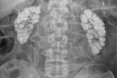
- Macroscopic nephrocalcinosis is visible on x-ray and ultrasound.
Deposits can accumulate in the internal medullary interstitium, in the basement membranes and in the bends of the loops of Henle, in the cortex and even in the lumens of the tubules. And depending on the localization, nephrocalcinosis is divided into medullary and cortical.
Medullary nephrocalcinosis is characterized by interstitial (intercellular) deposition of calcium oxalate and phosphate in the renal medulla - around the renal pyramids.
If the tissues of the cortical layer of the renal parenchyma undergo calcification - in the form of one or two stripes of deposited Ca salts along the zone of damaged cortical tissue or many small deposits scattered in the renal cortex - then cortical nephrocalcinosis is determined.
Diagnostics nephrocalcinosis
Diagnosis of nephrocalcinosis requires a complete examination of the kidneys. The pathology is usually diagnosed using visualization methods, since only instrumental diagnostics can detect Ca deposits using: ultrasound of the kidneys and ureters; [ 15 ] ultrasound Dopplerography of the kidneys, CT or MRI.
If the results of radiological methods in diagnosing the kidneys are not sufficiently convincing,a kidney biopsy may be required to confirm nephrocalcinosis.
Necessary laboratory tests include urine tests: general, Zimnitsky test, total calcium in urine, as well as excretion of phosphate, oxalate, citrate and creatinine. Tests are taken for total and ionized calcium in the blood, for the content of alkaline phosphase, parathyroid hormone, calcitonin in the blood plasma.
Given the multifactorial etiology of renal calcification, the range of diagnostic tests can be much wider. For example, to detect osteoporosis, CT of spongy bone tissue, X-ray absorptiometry and ultrasound densitometry are performed; more tests are required to determine MEN type I syndrome, etc. [ 16 ]
Differential diagnosis
Differential diagnosis is mandatory: with papillary necrosis of the kidney, tuberculosis of the kidney, pneumocystis or mycobacterial extrapulmonary infection in patients with AIDS, etc.
Nephrocalcinosis should not be confused with nephrolithiasis, that is, the formation of kidney stones, although in some patients both pathologies are present simultaneously.
Who to contact?
Treatment nephrocalcinosis
In the case of nephrocalcinosis, treatment is aimed not only at the etiologically related pathology and reducing existing symptoms, but also at reducing the calcium content in the blood.
Treatment of hypercalcemia requires an increase in fluid intake and hydration with isotonic sodium chloride solution, and calcimimetics (Cinacalcet, etc.) are also taken.
Therapy for macroscopic nephrocalcinosis may include thiazide diuretics and citrate drugs (potassium citrate, etc.) that promote better solubility of calcium in urine.
If nephrocalcinosis is associated with osteoporosis and increased bone resorption, antiresorptive agents (bone resorption inhibitors) are used – bisphosphonates and other drugs for the treatment and prevention of osteoporosis.
Phosphate-binding drugs (Sevelamer or Renvela) are prescribed for the treatment of hyperphosphatemia. Drug therapy for hypoaldosteronism is carried out with mineralocorticoids (Trimethylacetate, Florinef, etc.).
Patients with chronic hypoparathyroidism may be given recombinant human parathyroid hormone (Teriparatide).
Nephrocalcinosis, which occurs in the renal cortex during its necrosis, requires systemic antibiotic therapy, restoration of water-electrolyte balance by intravenous fluid administration and hemodialysis.
In order to limit the intake of certain macronutrients, a diet for nephrocalcinosis is prescribed, more details:
Surgical treatment does not involve removing calcium deposits from kidney tissue: only formed stones can be removed. Surgical intervention is possible in primary hyperparathyroidism, since by removing the parathyroid gland it is possible to stop the negative impact of its hormone on the kidneys. [ 17 ]
Prevention
Among the measures that can prevent the development of hypercalcemic nephropathy, experts name drinking enough water and reducing the consumption of salt and foods containing a lot of calcium and oxalates.
And, of course, timely treatment of diseases that lead to nephrocalcinosis.
Forecast
The etiology of calcium deposition in the kidneys and the nature of the complications of this process will determine the prognosis of nephrocalcinosis in each specific case. Specific causes of nephrocalcinosis, such as Dent's disease, primary hyperoxaluria and hypomagnesemic hypercalciuric nephrocalcinosis, in the absence of effective treatment can progress to chronic renal failure, [ 18 ] end-stage renal failure. Radiologically detected nephrocalcinosis is rarely reversible. The pathology rarely progresses, but medicine is not yet able to reduce the degree of calcification.