Medical expert of the article

New publications
Examination of cranial nerves. VII pair: facial nerve (n. facialis)
Last reviewed: 04.07.2025

All iLive content is medically reviewed or fact checked to ensure as much factual accuracy as possible.
We have strict sourcing guidelines and only link to reputable media sites, academic research institutions and, whenever possible, medically peer reviewed studies. Note that the numbers in parentheses ([1], [2], etc.) are clickable links to these studies.
If you feel that any of our content is inaccurate, out-of-date, or otherwise questionable, please select it and press Ctrl + Enter.
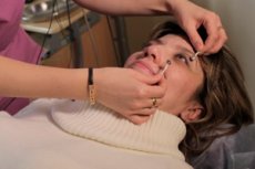
The examination of facial nerve functions begins with an assessment of the symmetry of the patient's face at rest and during spontaneous facial expressions. Particular attention is paid to the symmetry of the nasolabial folds and eye slits.
The motor fibers of the facial nerve innervate the facial muscles, the subcutaneous muscle of the neck (platysma), the stylohyoid, occipital muscles, the posterior belly of the digastric muscle, and the stapedius muscle. The autonomic parasympathetic fibers innervate the lacrimal gland, the sublingual and submandibular salivary glands, as well as the glands of the mucous membrane of the nose, hard and soft palate. The sensory fibers conduct taste impulses from the anterior two-thirds of the tongue and from the hard and soft palate.
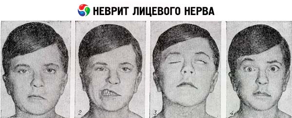
The strength of the facial muscles is tested one by one, asking the patient to wrinkle his forehead (m. frontalis), close his eyes tightly (m. orbicularis oculi), puff out his cheeks (m. buccinator), smile, show his teeth (m. risorius and m. zygomaticus major), purse his lips and not let them open (m. orbicularis oris). The patient is asked to take a breath and puff out his cheeks; normally, when pressing on the cheeks, the patient holds the air without releasing it through the mouth. If weakness of the facial muscles is detected, it is determined whether it concerns only the lower part of the face or extends to its entire half (both lower and upper).
The taste is tested on the front third of the tongue. The patient is asked to stick out the tongue and hold it by the tip with a gauze napkin. Using a pipette, drops of sweet, salty, and neutral solutions are applied to the tongue one by one. The patient must report the taste of the solution, pointing to the corresponding inscription on a piece of paper. It is noted whether tears are released when taste stimuli are applied (this paradoxical reflex is observed in patients with abnormal germination of secretory fibers after previous damage to the branches of the facial nerve).
The facial nerve contains a very small number of fibers that conduct impulses of general sensitivity and innervate small areas of the skin, one of which is located on the inner surface of the auricle near the external auditory canal, and the second - directly behind the ear. Pain sensitivity is examined by applying pin pricks directly behind the external auditory canal.
Signs of facial nerve damage
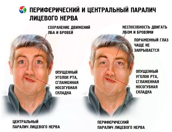
Damage to the central motor neuron (for example, in a hemispheric stroke ) can cause central, or "supranuclear", paralysis of the facial muscles. It is characterized by contralateral paresis of the facial muscles located only in the lower half of the face (very slight weakness of the orbicularis oculi muscle and slight asymmetry of the eye slits are possible, but the ability to wrinkle the forehead remains). This is explained by the fact that the part of the motor nucleus n. facialis that innervates the lower facial muscles receives impulses only from the opposite hemisphere, while the part that innervates the upper facial muscles is under the influence of the corticonuclear tracts of both hemispheres. Damage to the peripheral motor neuron (neurons of the motor nucleus n. facialis and their axons) results in peripheral paralysis of the facial muscles (prosoplegia), which is characterized by weakness of the facial muscles of the entire ipsilateral half of the face. Closure of the eyelids on the affected side is impossible ( lagophthalmos ) or is incomplete.
Bell's sign is often observed in patients with peripheral paralysis of the facial muscles: when the patient tries to close his eyes, the eyelids on the side of the facial nerve lesion do not close, and the eyeball moves upward and outward. The movement of the eyeball in this case is a physiological synkinesis, which consists in the movement of the eyeballs upward when closing the eyes. To see it in a healthy person, it is necessary to forcibly hold his eyelids in a raised position, asking him to close his eyes. Peripheral paralysis of the facial muscles in some cases can be accompanied by taste disturbances in the anterior two-thirds of the ipsilateral half of the tongue (with damage to the trunk of the facial nerve above the origin of the chorda tympani fibers from its distal part). With central paralysis of the facial muscles, that is, with damage to the corticonuclear tracts going to the motor nucleus of the facial nerve, taste disturbances do not occur.
Read also: Facial nerve paralysis
If the facial nerve is damaged above the point where its fibers branch off to the stapedius muscle, a distortion of the timbre of perceived sounds occurs - hyperacusis. If the facial nerve is damaged at the level of its exit from the pyramid of the temporal bone through the stylomastoid opening, the parasympathetic fibers to the lacrimal gland (n. petrosus major) and the sensory fibers coming from the taste buds (chorda tympani) do not suffer, so taste and lacrimation remain intact. Lacrimation on the side of lagophthalmos is characteristic, which is explained by excessive irritation of the mucous membrane of the eye due to the absence of a protective blink reflex and difficulty moving tears to the lower lacrimal canal due to sagging of the lower eyelid. All this leads to tears freely flowing down the face.
Bilateral acute or subacute lesion of the facial nerve of the peripheral type is observed in Guillain-Barré syndrome (GBS). Acute or subacute unilateral peripheral paralysis of the facial muscles most often occurs with compression-ischemic neuropathy of the facial nerve (with compression-ischemic changes in the section of the nerve that passes through the facial canal in the pyramid of the temporal bone.
During the recovery period after peripheral paralysis, pathological regeneration of facial nerve fibers is possible.
In this case, on the side of paralysis, over time, contracture of the facial muscles develops, due to which the palpebral fissure becomes narrower, and the nasolabial fold becomes deeper than on the healthy side (the face is "skewed" not to the healthy, but to the diseased side). Contracture of the facial muscles usually occurs against the background of residual phenomena of prosoparesis and is combined with pathological synkinesis of the facial muscles. For example, when squinting the eyes on the diseased side, the corner of the mouth involuntarily rises (labio-periorbital synkinesis), or the wing of the nose rises, or the platysma contracts; when puffing out the cheeks, the palpebral fissure narrows, etc.