Medical expert of the article

New publications
Examination of cranial nerves. Pair II: optic nerve (n. opticus)
Last reviewed: 07.07.2025

All iLive content is medically reviewed or fact checked to ensure as much factual accuracy as possible.
We have strict sourcing guidelines and only link to reputable media sites, academic research institutions and, whenever possible, medically peer reviewed studies. Note that the numbers in parentheses ([1], [2], etc.) are clickable links to these studies.
If you feel that any of our content is inaccurate, out-of-date, or otherwise questionable, please select it and press Ctrl + Enter.
The optic nerve conducts visual impulses from the retina of the eye to the cortex of the occipital lobe.
When collecting anamnesis, it is determined whether the patient has any changes in vision. Changes in visual acuity (distant or near) are the responsibility of an ophthalmologist. In case of transient episodes of blurred vision, limited visual fields, photopsies or complex visual hallucinations, a detailed examination of the entire visual analyzer is necessary. The most common cause of transient visual impairment is migraine with visual aura. Visual disturbances are most often represented by flashes of light or sparkling zigzags (photopsies), flickering, loss of a section or the entire visual field. The visual aura of migraine develops 0.5-1 hour (or less) before the headache attack, lasts on average 10-30 minutes (no more than 1 hour). Headache with migraine occurs no later than 60 minutes after the end of the aura. Visual hallucinations such as photopsies (flashes, sparks, zigzags) may represent the aura of an epileptic seizure in the presence of a pathological focus irritating the cortex in the area of the calcarine groove.

Visual acuity and its study
Visual acuity is determined by ophthalmologists. To assess visual acuity at a distance, special tables with circles, letters, and numbers are used. The standard table used in Ukraine contains 10-12 rows of signs (optotypes), the sizes of which decrease from top to bottom in an arithmetic progression. Vision is examined from a distance of 5 m, the table should be well lit. The norm (visual acuity 1) is such visual acuity at which the subject is able to distinguish the optotypes of the 10th (counting from the top) line from this distance. If the subject is able to distinguish the signs of the 9th line, his visual acuity is 0.9, the 8th line - 0.8, etc. In other words, reading each subsequent line from top to bottom indicates an increase in visual acuity by 0.1. Near visual acuity is checked using other special tables or by asking the patient to read text from a newspaper (normally, small newspaper print can be distinguished from a distance of 80 cm). If visual acuity is so poor that the patient cannot read anything from any distance, they limit themselves to counting fingers (the doctor's hand is positioned at the level of the patient's eyes). If this is also impossible, the patient is asked to determine whether he is in a dark or lit room. Reduced visual acuity ( amblyopia ) or complete blindness (amaurosis) occurs when the retina or optic nerve is damaged. With such blindness, the direct reaction of the pupil to light disappears (due to the interruption of the afferent part of the pupillary reflex arc), but the reaction of the pupil in response to illumination of the healthy eye remains intact (the efferent part of the pupillary reflex arc, represented by the fibers of the third cranial nerve, remains intact). Slowly progressive vision loss is observed when the optic nerve or chiasm is compressed by a tumor.
Signs of violations
Transient short-term loss of vision in one eye (transient monocular blindness, or amaurosis fugax - from the Latin "fleeting") can be caused by a transient disruption of the blood supply to the retina. It is described by the patient as a "curtain falling from top to bottom" when it occurs and as a "rising curtain" when it reverses. Vision is usually restored within a few seconds or minutes. Acute and progressive reduction in vision over 3-4 days, then restored within a few days to weeks and often accompanied by eye pain, is characteristic of retrobulbar neuritis. Sudden and persistent loss of vision occurs with fractures of the bones of the anterior cranial fossa in the area of the optic canal; with vascular lesions of the optic nerve and temporal arteritis. When the bifurcation zone of the basilar artery is blocked and a bilateral infarction of the occipital lobes develops with damage to the primary visual centers of both cerebral hemispheres, "tubular" vision or cortical blindness occurs. "Tubular" vision is caused by bilateral hemianopsia with preservation of central (macular) vision in both eyes. Preservation of vision in a narrow central visual field is explained by the fact that the macular projection zone at the pole of the occipital lobe is supplied with blood from several arterial basins and, in case of infarction of the occipital lobes, most often remains intact. Visual acuity in these patients is slightly reduced, but they behave as if they were blind. "Cortical" blindness occurs in the case of insufficiency of anastomoses between the cortical branches of the middle and posterior cerebral arteries in the areas of the occipital cortex responsible for central (macular) vision. Cortical blindness is characterized by the preservation of pupillary reactions to light, since the visual pathways from the retina to the brainstem are not damaged. Cortical blindness with bilateral damage to the occipital lobes and parieto-occipital areas in some cases may be combined with denial of this disorder, achromatopsia, apraxia of conjugate eye movements (the patient cannot direct his gaze toward an object located in the peripheral part of the visual field) and the inability to visually perceive an object and touch it. The combination of these disorders is called Balint's syndrome.
Fields of vision and their study
The visual field is the area of space that a motionless eye sees. The integrity of the visual fields is determined by the state of the entire visual pathway (optic nerves, optic tract, optic radiation, cortical visual area, which is located in the calcarine groove on the medial surface of the occipital lobe). Due to the refraction and crossing of light rays in the lens and the transition of visual fibers from the same halves of the retina in the chiasm, the right half of the brain is responsible for the integrity of the left half of the visual field of each eye. Visual fields are assessed separately for each eye. There are several methods for their approximate assessment.
- Alternate assessment of individual visual fields. The doctor sits opposite the patient. The patient covers one eye with his palm and looks at the bridge of the doctor's nose with the other eye. A hammer or wiggling fingers are moved along the perimeter from behind the patient's head to the center of his visual field and the patient is asked to note the moment the hammer or fingers appear. The examination is conducted alternately in all four quadrants of the visual fields.
- The "threat" method is used in cases where it is necessary to examine the visual fields of a patient who is inaccessible to speech contact (aphasia, mutism, etc.). The doctor, with a sharp "threatening" movement (from the periphery to the center), brings the extended fingers of his hand closer to the patient's pupil, observing its blinking. If the visual field is intact, the patient blinks in response to the finger approaching. All visual fields of each eye are examined.
The described methods are related to screening; visual field defects are more accurately detected using a special device - a perimeter.
Signs of violations
Monocular visual field defects are usually caused by pathology of the eyeball, retina or optic nerve - in other words, damage to the visual pathways before their crossing (chiasm) causes a visual field disorder in only one eye, located on the affected side. Binocular visual field defects (hemianopsia) can be bitemporal (both eyes have temporal visual fields loss, i.e. the right eye has the right, the left eye has the left) or homonymous (each eye has the same visual fields loss - either left or right). Bitemporal visual field defects occur with lesions in the area of the crossing of optic fibers (for example, damage to the chiasm in onyxoj and the pituitary gland). Homonymous visual field defects occur when the optic tract, optic radiation, or visual cortex are affected, i.e. when the visual pathway above the chiasm is affected (these defects occur in the visual fields opposite to the lesion: if the lesion is in the left hemisphere, the right visual fields of both eyes are affected, and vice versa). Damage to the temporal lobe results in defects in the homonymous upper quadrants of the visual fields (contralateral upper quadrant anopsia), and damage to the parietal lobe results in defects in the homonymous lower quadrants of the visual fields (contralateral lower quadrant anopsia).
Conduction visual field defects are rarely associated with changes in visual acuity. Even with significant peripheral visual field defects, central vision may be preserved. Patients with visual field defects caused by damage to the visual pathways above the chiasm may not be aware of their presence, especially in cases of parietal lobe damage.
 [ 1 ]
[ 1 ]
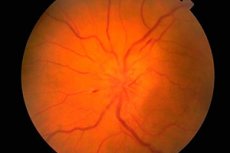
The fundus and its examination
The fundus is examined using an ophthalmoscope. The condition of the optic nerve head (papilla) (the initial, intraocular part of the optic nerve visible during ophthalmoscopy), the retina, and the vessels of the fundus are assessed. The most important characteristics of the fundus are the color of the optic nerve head, the clarity of its borders, the number of arteries and veins (usually 16-22), the presence of venous pulsation, any abnormalities or pathological changes: hemorrhages, exudate, changes in the walls of blood vessels in the area of the yellow spot (macula) and on the periphery of the retina.
Signs of violations
Edema of the optic disc is characterized by its bulging (the disc protrudes above the level of the retina and protrudes into the cavity of the eyeball), redness (the vessels on the disc are sharply dilated and filled with blood); the boundaries of the disc become unclear, the number of retinal vessels increases (more than 22), the veins do not pulsate, hemorrhages are present. Bilateral edema of the optic disc ( congestive papilla of the optic nerve ) is observed with increased intracranial pressure (volume process in the cranial cavity, hypertensive encephalopathy, etc.). Visual acuity is initially usually not affected. If the increase in intracranial pressure is not eliminated in a timely manner, visual acuity gradually decreases and blindness develops due to secondary atrophy of the optic nerve.
Congestion of the optic nerve head must be differentiated from inflammatory changes (papillitis, optic neuritis ) and ischemic optic neuropathy. In these cases, the changes in the head are often unilateral, pain in the eyeball area and decreased visual acuity are typical. Paleness of the optic nerve head in combination with decreased visual acuity, narrowing of the visual fields, decreased pupillary reactions are characteristic of optic nerve atrophy, which develops in many diseases affecting this nerve (inflammatory, dysmetabolic, hereditary). Primary optic nerve atrophy develops with damage to the optic nerve or chiasm, while the head is pale, but has clear boundaries. Secondary optic nerve atrophy develops following edema of the optic nerve head, the boundaries of the head are initially unclear. Selective pallor of the temporal half of the optic disc may be observed in multiple sclerosis, but this pathology can easily be confused with a variant of the normal state of the optic disc. Pigmentary degeneration of the retina is possible in degenerative or inflammatory diseases of the nervous system. Other important pathological findings for a neurologist during examination of the fundus include arteriovenous angioma of the retina and the cherry pit symptom, which is possible in many gangliosidoses and is characterized by the presence of a white or gray rounded lesion in the macula, in the center of which there is a cherry-red spot. Its origin is associated with atrophy of the retinal ganglion cells and the translucency of the vascular membrane through it.