Medical expert of the article

New publications
Suboccipital muscles
Last reviewed: 07.07.2025

All iLive content is medically reviewed or fact checked to ensure as much factual accuracy as possible.
We have strict sourcing guidelines and only link to reputable media sites, academic research institutions and, whenever possible, medically peer reviewed studies. Note that the numbers in parentheses ([1], [2], etc.) are clickable links to these studies.
If you feel that any of our content is inaccurate, out-of-date, or otherwise questionable, please select it and press Ctrl + Enter.
The suboccipital muscles (mm. suboccipitales) include the rectus capitis posterior major, rectus capitis posterior minor, and the superior and inferior oblique muscles of the capitis. These muscles are located deep under the semispinalis, longissimus, and splenius capitis muscles. The suboccipital muscles border the suboccipital triangular space (trigonum suboccipitile), which contains the vertebral artery, the posterior branch of the first cervical spinal nerve, the posterior arch of the atlas, and the posterior atlantooccipital membrane.
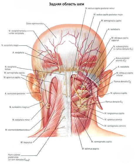
The large posterior rectus capitis muscle (m. rectus capitis posterior major) originates on the spinous process of the axial vertebra and is attached to the occipital bone under the inferior nuchal line.
Function: throws the head back, tilts it to the side, and with a one-sided contraction turns the head to one side.
Innervation: suboccipital nerve.
Blood supply: deep cervical artery.
The small posterior rectus capitis muscle (m. rectus capitis posterior minor) originates on the posterior tubercle of the atlas and is attached to the occipital bone under the inferior nuchal line, deeper and medial to the large posterior rectus capitis muscle.
Function: throws back and tilts the head to the side.
Innervation: suboccipital nerve (CI).
Blood supply: deep cervical artery.
The inferior oblique muscle of the head (m. obliquus capitis inferior) begins on the spinous process of the axial vertebra, passes upward and laterally, and attaches to the transverse process of the atlas.
Function: extends, tilts to the side and rotates the head around the longitudinal axis of the axial vertebra.
Innervation: suboccipital nerve (CI).
Blood supply: deep cervical artery.
The superior oblique muscle of the head (m. obliquus capitis superior) originates on the transverse process of the atlas, passes upward and medially, and is attached to the occipital bone above the inferior nuchal line. The muscle lies deeper and lateral to the attachment of the semispinalis capitis muscle.
Function: with bilateral contraction, the muscle extends the head; with unilateral contraction, it tilts the head laterally to its side.
Innervation: suboccipital nerve (CI).
Blood supply: deep cervical artery.
 [
[ What do need to examine?
How to examine?