Medical expert of the article

New publications
Back muscles
Last reviewed: 04.07.2025

All iLive content is medically reviewed or fact checked to ensure as much factual accuracy as possible.
We have strict sourcing guidelines and only link to reputable media sites, academic research institutions and, whenever possible, medically peer reviewed studies. Note that the numbers in parentheses ([1], [2], etc.) are clickable links to these studies.
If you feel that any of our content is inaccurate, out-of-date, or otherwise questionable, please select it and press Ctrl + Enter.
The back muscles (musculi dorsi) are paired and occupy the entire dorsal side of the body, starting from the sacrum and adjacent parts of the iliac crests to the base of the skull. Arranged in layers, these muscles have complex anatomical and topographic relationships due to the peculiarities of their development and function. There are superficial and deep muscles of the back. The muscles are covered with fascia, which separate one group of muscles from another.
Most of the superficial muscles of the back develop in connection with the upper limb. These include the trapezius, latissimus dorsi, levator scapulae, rhomboid major and minor. The serratus posterior superior and inferior are deeper and attach to the ribs.
The deep muscles, which make up the majority of the back muscles, are derivatives of myotomes - muscle rudiments of the primary body segments - somites. These muscles include the strap muscles of the head and neck, the muscle that straightens the trunk, the suboccipital muscles, etc.
 [ 1 ]
[ 1 ]
Superficial muscles of the back
The superficial muscles of the back are attached to the bones of the shoulder girdle and humerus and are arranged in two layers. The first layer is formed by the trapezius muscle and the latissimus dorsi muscle, the second - by the large and small rhomboid muscles, the muscle that lifts the scapula, the upper and lower serratus muscles.
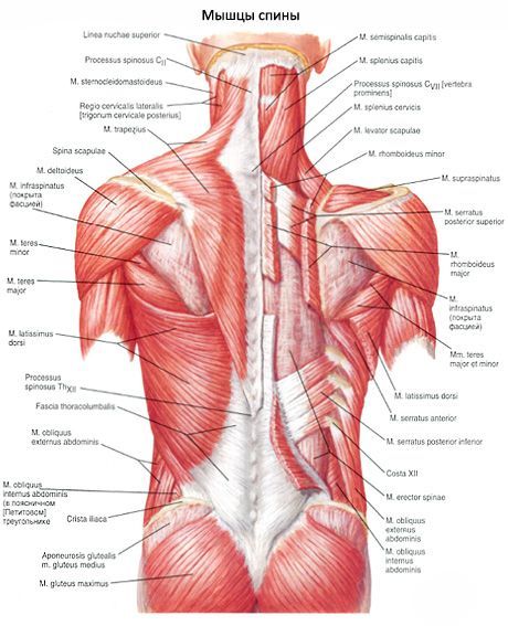
The trapezius muscle (m. trapezius) is flat, triangular in shape, with a wide base facing the posterior midline. The muscle occupies the upper part of the back and the back of the neck.
The latissimus dorsi muscle (m. latissimus dorsi) is flat, triangular in shape, and occupies the lower half of the back on the corresponding side. The latissimus dorsi muscle lies superficially, with the exception of the upper edge, which is hidden under the lower part of the trapezius muscle. Below, the lateral edge of the latissimus dorsi muscle forms the medial side of the lumbar triangle (the lateral side of this triangle is formed by the edge of the external oblique abdominal muscle, the lower side by the iliac crest).
The muscle that raises the scapula (m. levator scapulae) begins with tendinous bundles on the posterior tubercles of the transverse processes of the upper three or four cervical vertebrae (between the attachment sites of the middle scalene muscle - in front and the splenius muscle of the neck - behind). Directing downwards, the muscle attaches to the medial edge of the scapula, between its upper angle and spine.
The small and large rhomboid muscles (mm. rhomboidei minor et major) often grow together and form one muscle. The small rhomboid muscle originates on the lower part of the nuchal ligament, the spinous processes of the 7th cervical and 1st thoracic vertebrae, and on the supraspinous ligament.
Rhomboid minor and major muscles
Two thin flat muscles are attached to the ribs - the upper and lower posterior serratus muscles.
Upper and lower serratus posterior muscles
Deep back muscles
The deep muscles of the back form three layers: superficial, middle and deep. The superficial layer is represented by the splenius capitis, splenius cervicis and erector spinae muscles. The middle layer is formed by the transverse spinal muscle. The deep layer is formed by the interspinous, intertransverse and suboccipital muscles.
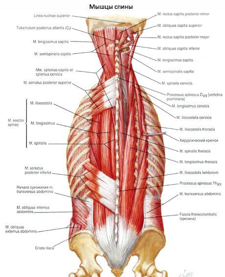
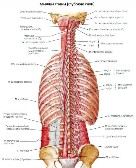
The muscles of the superficial layer are the most developed, belonging to the type of strong muscles that perform mainly static work. They extend along the entire length of the back and the back of the neck from the sacrum to the occipital bone. The origin and attachment sites of these muscles occupy vast surfaces. Therefore, when they contract, the muscles of the superficial layer develop great force, holding the spine in a vertical position, which serves as a support for the head, ribs, viscera and upper limbs. The muscles of the middle layer are located obliquely, and are thrown from the transverse to the spinous processes of the vertebrae. They form several layers, and in the deepest layer, the muscle bundles are the shortest and are attached to adjacent vertebrae. The more superficial the muscle bundles are, the longer they are and the more vertebrae they are thrown over (from 5 to 6). In the deepest, third layer, the short muscles are not located at all levels of the spine. These muscles are well developed in the most mobile sections of the spinal column: cervical, lumbar and lower thoracic. The third layer also includes muscles that act on the atlanto-occipital joint. These muscles are called suboccipital muscles (mm. suboccipitals).
The deep muscles of the back become visible after the superficial muscles have been dissected and cut layer by layer: the latissimus dorsi, trapezius, rhomboid and serratus muscles.
The splenitis capitis muscle (m. splenitis capitis) is located in front of the upper part of the sternocleidomastoid and trapezius muscles. It begins on the lower half of the nuchal ligament (below the level of the fourth cervical vertebra), on the spinous processes of the seventh cervical and upper three to four thoracic vertebrae. The bundles of this muscle go upward and laterally and are attached to the mastoid process of the temporal bone and to the area under the lateral part of the superior nuchal line of the occipital bone.
The splenius cervicis muscle (m. splenius cervicis) originates on the spinous processes of the III-IV thoracic vertebrae. It is attached to the posterior tubercles of the transverse processes of the two or three upper cervical vertebrae. The muscle covers the beginning of the bundles of the muscle that lifts the scapula from behind. Behind it is the trapezius muscle.
The erector spinae muscle is the strongest of the autochthonous muscles of the back, extending along the entire length of the spine - from the sacrum to the base of the skull. It lies in front of the trapezius, rhomboid, posterior serratus muscles, and the latissimus dorsi. Behind, the erector spinae muscle is covered by the superficial layer of the lumbosacral fascia.
The iliocostalis muscle (m. iliocostalis) is the most lateral part of the muscle that straightens the spine. This muscle begins on the iliac crest, the inner surface of the superficial leaflet of the lumbosacral fascia. Muscle bundles pass upward along the back surface of the ribs laterally from their angles to the transverse processes of the lower (VII-IV) cervical vertebrae. According to the location of the individual parts of the muscle, it is divided into the iliocostalis lumborum muscle, the iliocostalis thoracic muscle, and the iliocostalis cervicis muscle.
The longissimus muscle (m. longissimus) is the largest of the three muscles that form the muscle that straightens the spine.
The spinal muscle (m. spinalis) is the most medial of the three parts of the muscle that straightens the spine. The muscle is directly adjacent to the spinous processes of the thoracic and cervical vertebrae. This muscle is divided into the spinal muscle of the chest, the spinal muscle of the neck, and the spinal muscle of the head.
The transverse spinal muscle (m. transversospinalis) is represented by a multitude of layered muscle bundles that pass obliquely upward from the lateral to the medial side from the transverse processes to the spinous processes of the vertebrae. The muscle bundles of the transverse spinal muscle have unequal lengths and, throwing over a different number of vertebrae, form individual muscles: semispinalis, multifidus, and rotator muscles.
The multifidus muscles (mm. multiridi) are muscle-tendon bundles that originate on the transverse processes of the underlying vertebrae and attach to the spinous processes of the overlying ones.
The rotator muscles of the neck, chest and lower back (mm. rotatores cervicis, thoracis et lumborum) are located in the deepest layer of the back muscles, in the groove between the spinous and transverse processes. These muscles are best expressed within the thoracic spine. According to the length of the bundles, they are divided into long and short.
Rotator muscles of the neck, chest and lower back
The muscles that raise the ribs (mm. levatores costarum) are divided into short and long. The short muscles occupy the posterior sections of the intercostal spaces medially from the external intercostal muscles.
The interspinous muscles of the neck, chest and lower back (mm. interspinales cervicis, thoracis et lumborum) connect the spinous processes of the vertebrae with each other, starting from the second cervical and below. They are better developed in the cervical and lumbar sections of the spinal column, which are characterized by the greatest mobility. In the thoracic part of the spine, the interspinous muscles are weakly expressed (may be absent).
Interspinous muscles of the neck, chest and lumbar region
The intertransverse muscles of the lumbar region, chest and neck (mm. intertransversarii lumborum, thoracis et cervicis) are formed by short bundles connecting the transverse processes of adjacent vertebrae, and are best expressed at the level of the lumbar and cervical spine. The intertransverse muscles of the lumbar region are subdivided into lateral and medial.
Intertransverse muscles of the lumbar, thoracic and cervical spine
The suboccipital muscles (mm. suboccipitales) include the rectus capitis posterior major, rectus capitis posterior minor, and the superior and inferior oblique muscles of the capitis. These muscles are located deep under the semispinalis, longissimus, and splenius capitis muscles. The suboccipital muscles border the suboccipital triangular space (trigonum suboccipitile), which contains the vertebral artery, the posterior branch of the first cervical spinal nerve, the posterior arch of the atlas, and the posterior atlantooccipital membrane.
[ 2 ]