Medical expert of the article

New publications
Chest muscles
Last reviewed: 04.07.2025

All iLive content is medically reviewed or fact checked to ensure as much factual accuracy as possible.
We have strict sourcing guidelines and only link to reputable media sites, academic research institutions and, whenever possible, medically peer reviewed studies. Note that the numbers in parentheses ([1], [2], etc.) are clickable links to these studies.
If you feel that any of our content is inaccurate, out-of-date, or otherwise questionable, please select it and press Ctrl + Enter.
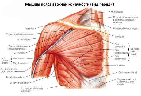
The chest muscles are arranged in several layers. More superficial are those muscles that develop in connection with the formation of the upper limb. They connect the upper limb to the chest. These include the pectoralis major and the anterior serratus muscles. Each muscle has its own fascia. In front of the superficial muscles is the superficial (subcutaneous) fascia of the chest.
The deep layers of the chest muscles are represented by their own, autochthonous muscles, developing from the ventral sections of the myotomes. These muscles begin and are attached within the chest wall. These include the external and internal intercostal muscles, the subcostal muscles, the transverse muscle of the chest, and the muscles that raise the ribs.
Together with the chest muscles, the closely related anatomically and functionally thoracoabdominal obstruction is described - the diaphragm - the main respiratory muscle, which develops from the ventral parts of the cervical myotomes.
 [
[ Superficial muscles of the chest
The pectoralis major muscle (m. pectoralis major) is massive, fan-shaped, and occupies a significant portion of the anterior wall of the chest cavity. According to the places of its origin, the muscle is divided into the clavicular part (pars clavicularis), which begins on the medial half of the clavicle; the sternocostal part (pars sternocostalis), which begins on the anterior surface of the sternum and the cartilages of the upper six ribs, and the abdominal part (pars abdominalis), which begins on the anterior wall of the sheath of the rectus abdominis muscle.
The pectoralis minor muscle (m. pectoralis minor) is flat, triangular in shape, and is located directly behind the pectoralis major muscle. The muscle begins on the II-V ribs, near their anterior ends. Directed upward and laterally, it is attached by a short tendon to the coracoid process of the scapula.
The subclavian muscle (m. subclavius) is small in size and occupies a slit-like space between the 1st rib and the clavicle. It begins on the cartilage of the 1st rib, runs laterally, and attaches to the lower surface of the acromial end of the clavicle.
The anterior serratus muscle (m. serratus anterior) is wide, quadrangular in shape, adjoins the rib cage from the side, forms the medial wall of the axillary cavity. It begins with large teeth on the upper eight to nine ribs and is attached to the medial edge and lower angle of the scapula. The upper and middle bundles of the muscle lie horizontally, the lower bundles are located obliquely and pass from front to back and from bottom to top.
Deep chest muscles
The external intercostal muscles (mm. intercostales externi), 11 on each side, begin at the lower edge of the overlying rib, outside of its groove, and, directed downwards and forwards, are attached to the upper edge of the underlying rib. The muscles occupy the intercostal spaces from the tubercles of the ribs at the back to the costal cartilages in front, where their continuation to the edge of the sternum is the external intercostal membrane (membrane - membrana intercostalis externa).
The internal intercostal muscles (mm. intercostales interni) are located medially from the external intercostal muscles. They occupy the intercostal spaces, starting from the edge of the sternum (at the true ribs) and the anterior ends of the cartilages of the false ribs and to the angles of the ribs at the back, where their continuation is the internal intercostal membrane (membrane - membrana intercostalis interna).
The subcostal muscles (mm. subcostales) are formed by muscle and tendon bundles in the lower part of the posterior section of the inner surface of the chest wall. They begin on the X-XII ribs near their angles, are directed upward and laterally, are thrown over one or two ribs and are attached to the inner surface of the overlying ribs.
The transverse thoracic muscle (m. transversus thoracis) is located on the back (inner) surface of the anterior chest wall. This muscle begins on the xiphoid process, the lower half of the body of the sternum.