Medical expert of the article

New publications
Muscle research
Last reviewed: 04.07.2025

All iLive content is medically reviewed or fact checked to ensure as much factual accuracy as possible.
We have strict sourcing guidelines and only link to reputable media sites, academic research institutions and, whenever possible, medically peer reviewed studies. Note that the numbers in parentheses ([1], [2], etc.) are clickable links to these studies.
If you feel that any of our content is inaccurate, out-of-date, or otherwise questionable, please select it and press Ctrl + Enter.
A detailed study of the muscular system, including the identification of various developmental disorders, tone, muscle strength, and functions of individual muscles, is usually carried out by a neurologist and is therefore studied in detail in the course on nervous diseases. However, a doctor of any specialty must master the basic techniques of studying the muscular system, since certain changes in it can also be encountered in diseases of internal organs.
Complaint assessment
First of all, the presence of complaints of the patient about muscle weakness and muscle fatigue when performing various movements is noted. Sometimes these complaints concern many muscle groups, but more often they affect very specific groups (for example, chewing, facial muscles, etc.). The patient may also complain of involuntary fibrillary twitching of individual muscle groups, limitation and complete absence of active (voluntary) movements.
 [ 1 ], [ 2 ], [ 3 ], [ 4 ], [ 5 ], [ 6 ], [ 7 ], [ 8 ], [ 9 ]
Inspection and palpation
During the examination, attention is primarily paid to the degree of development of muscle tissue, the presence of atrophy or hypertrophy of individual muscles and muscle groups. Muscle atrophy is often observed in patients with peripheral paralysis and paresis, with spinal cord injury, prolonged forced stay in a motionless position (the so-called atrophy from inactivity). In the presence of atrophy of individual muscles or asymmetry in their development, the circumference of the shin, thigh, shoulder, forearm on the healthy side and on the affected side is measured and compared. Muscle hypertrophy is much less common (for example, in some hereditary diseases) and usually concerns individual muscle groups (gastrocnemius, quadriceps, deltoid).
When palpating individual muscles, pain may be detected (for example, in myositis). By directly palpating the muscles of symmetrical areas of the body, muscle tone is also determined, changes in which in some cases have great diagnostic value. When muscle tone decreases (hypotonia), muscle tissue appears soft, flabby, doughy. When muscle tone increases (hypertonicity), muscle tissue becomes, on the contrary, denser than normal.
[ 10 ], [ 11 ], [ 12 ], [ 13 ], [ 14 ], [ 15 ]
Assessment of muscle tone and strength
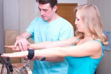
Some special techniques are also used to assess muscle tone. After asking the patient not to resist, the doctor himself makes passive movements (flexion and extension) of the patient's limbs in the shoulder, elbow and wrist joints. With the patient in a horizontal position on his back, the same movements are made in the hip, knee and ankle joints. In this case, the muscle tone of the right and left limbs is necessarily compared. With a decrease in muscle tone, passive flexion and extension of the corresponding limb occurs unusually easily, in the absence of the slight resistance that normally exists. With hypertonicity, muscle resistance is, on the contrary, increased. By raising and lowering the patient's head, it is possible to assess the tone of the neck muscles. A decrease in the tone of these muscles is easily detected if, having raised the patient's head, you suddenly take your hand away from it. Muscle tone is determined more accurately using special devices (myotonometers).
Muscle strength is assessed by the resistance that the patient is able to overcome. The doctor asks the patient to bend his arm at the elbow joint, and then, asking the patient to resist, tries to straighten it. In the same way, the patient's muscle strength can be tested by asking him to bend his leg at the knee joint, his hand at the wrist joint, his foot at the ankle joint, etc. When examining the muscle strength of the shoulder extensors, the doctor tries to bend the patient's arm at the elbow joint, which the patient holds in an extended position. It is clear that the study is carried out separately for the muscles of the right and left limbs.
Muscle strength is assessed on a five-point (sometimes six-point) system. In this case, in the case of normal muscle strength, the highest points are given, and in the case of its complete absence, the lowest (0). For a more accurate determination of muscle strength, special dynamometers are used.
One of the indicators of muscle strength is muscle fatigue. It is quite easy to detect if you ask the patient to quickly clench and unclench his fingers into a fist several times in a row. You can also ask the patient to stretch both arms forward. If muscle fatigue is present, the patient's arms (or one of them) quickly drop.
When examining the muscular system, attention is paid to the presence of another type of movement disorder - violent movements ( hyperkinesis ), which can occur in patients with rheumatism ( rheumatic chorea ), alcoholism, Parkinson's disease, and sometimes in elderly and senile people (senile tremor). In addition, in some diseases, involuntary muscle contractions are also observed, called cramps. It is customary to distinguish between clonic cramps, when muscle contractions are replaced by distinct periods of their relaxation, and tonic cramps, in which spastic muscle contractions occur, and periods of relaxation are expressed very weakly and are practically not noticeable.
How to examine?
What tests are needed?