Medical expert of the article

New publications
The sky
Last reviewed: 04.07.2025

All iLive content is medically reviewed or fact checked to ensure as much factual accuracy as possible.
We have strict sourcing guidelines and only link to reputable media sites, academic research institutions and, whenever possible, medically peer reviewed studies. Note that the numbers in parentheses ([1], [2], etc.) are clickable links to these studies.
If you feel that any of our content is inaccurate, out-of-date, or otherwise questionable, please select it and press Ctrl + Enter.
The palate (palatum) is divided into hard and soft. The bony basis of the hard palate (palatum durum) is made up of the palatine processes of the maxillary bones connected to each other, to which the horizontal plates of the palatine bones are attached at the back.
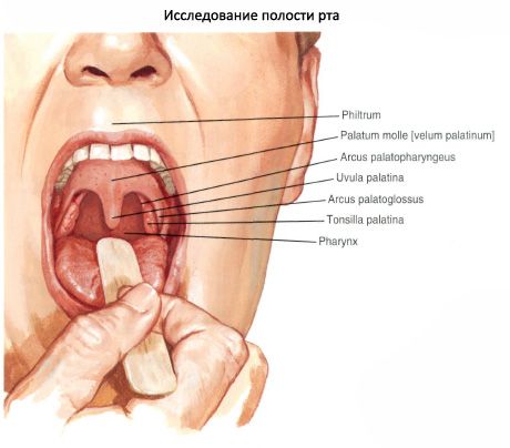
The soft palate (palatum molle) is attached to the posterior edge of the hard palate. The soft palate is based on a connective tissue plate (palatine aponeurosis) and the muscles of the soft palate, covered with a mucous membrane on the side of the nasal and oral cavities. The anterior part of the soft palate is located in the horizontal plane, the posterior, freely hanging edge of the palate is called the soft palate (velum palatinum). On the free edge of the soft palate there is a rounded process - the uvula (uvula palatina). Two folds (arches) begin from the lateral edges of the soft palate. The palatoglossus arch (Arcus palatoglossus) goes down to the lateral edge of the root of the tongue. The posterior, palatopharyngeal arch (arcus palatopharyngeus) goes down to the lateral wall of the pharynx. Between the arches is the tonsil fossa (fossa tonsillaris). It contains an organ of the immune system - the palatine tonsil (tonsilla palatina).
Paired striated muscles participate in the formation of the soft palate.
The muscle that tenses the soft palate (m.tensor veli palatini) begins on the cartilaginous part of the auditory tube, on the spine of the sphenoid bone. The muscle then goes down, bends around the pterygoid hook, is directed medially and is woven into the palatine aponeurosis. When contracting, the muscle pulls the soft palate and widens the lumen of the auditory tube.
The muscle that raises the soft palate (m.levator veli palatini) originates on the anterior half of the lower surface of the pyramid of the temporal bone and on the cartilaginous part of the auditory tube. This muscle passes medially to the previous muscle and is woven from above into the palatine aponeurosis. When this muscle contracts, the soft palate rises.
The uvula muscle (m.uvulae) begins on the posterior nasal spine and ends in the thickness of the mucous membrane of the uvula. When contracted, the muscle lifts and shortens the uvula.
The palatoglossus muscle (m.palatoglossus) begins in the lateral part of the root of the tongue, goes up in the thickness of the arch of the same name and is attached to the palatine aponeurosis. When contracted, the muscle lowers the soft palate, reducing the size of the pharynx.
The palatopharyngeal muscle (m.palatopharyngeus) begins in the thickness of the posterior wall of the pharynx and on the posterior edge of the plate of the cricoid cartilage, and is woven into the palatine aponeurosis. The muscle lowers the soft palate, reducing the size of the pharynx.
What do need to examine?