Medical expert of the article

New publications
Female genitalia
Last reviewed: 04.07.2025

All iLive content is medically reviewed or fact checked to ensure as much factual accuracy as possible.
We have strict sourcing guidelines and only link to reputable media sites, academic research institutions and, whenever possible, medically peer reviewed studies. Note that the numbers in parentheses ([1], [2], etc.) are clickable links to these studies.
If you feel that any of our content is inaccurate, out-of-date, or otherwise questionable, please select it and press Ctrl + Enter.
Internal female genital organs
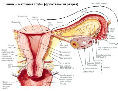
Ovary
The ovary (ovarium; Greek oophoron) is a paired organ, a female sex gland, located in the pelvic cavity behind the broad ligament of the uterus. In the ovaries, female sex cells (eggs) develop and mature, and female sex hormones are formed that enter the blood and lymph. The ovary has an ovoid shape, somewhat flattened in the anteroposterior direction.
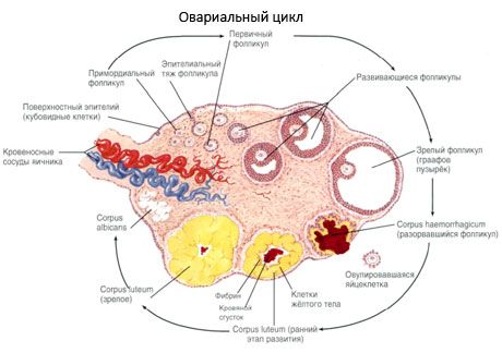
Oogenesis
Unlike male reproductive cells, egg cells multiply, their number increases in embryos, female fetuses, i.e. when the fetus is still in the mother's womb. In this case, so-called primordial follicles are formed, located in the deep layers of the ovarian cortex. Each such primordial follicle contains a young female reproductive cell - oogonia, surrounded by one layer of follicular cells.
Epididymis
Near each ovary there is a rudimentary formation - an ovarian appendage, a parovarian appendage (an appendage of the appendage), vesicular appendages, and the remains of the tubules of the primary kidney and its duct.
Uterus
The uterus (Greek metra) is an unpaired hollow muscular organ in which the embryo develops and the fetus is carried. The uterus is located in the middle part of the pelvic cavity behind the bladder and in front of the rectum. The uterus is pear-shaped, flattened in the anteroposterior direction. The uterus has a fundus, body and neck.
Placenta
The placenta, or baby's place, is a temporary organ that forms in the mucous membrane during pregnancy and connects the fetus's body with the mother's. The placenta is used to nourish the fetus, supply it with oxygen, and remove metabolic waste products from the fetus's body. The placenta protects the fetus's body from harmful substances (protective, barrier function). The blood of the mother and fetus in the placenta does not mix due to the presence of the so-called hematoplacental barrier.
Fallopian tube
The fallopian tube (tuba uterina, s.salpinx) is a paired organ used to conduct the egg from the ovary (from the peritoneal cavity) to the uterine cavity. The fallopian tubes are located in the pelvic cavity and are cylindrical ducts that run from the uterus to the ovaries. Each tube is located in the upper part of the broad ligament of the uterus, which is like a mesentery of the fallopian tube.
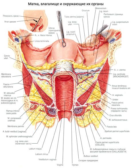
Vagina
The vagina (vagina, s.colpos) is an unpaired hollow organ shaped like a tube, located in the pelvic cavity and extending from the uterus to the genital slit. At the bottom of the vagina it passes through the urogenital diaphragm. The length of the vagina is 8-10 cm, the thickness of the wall is about 3 mm. The vagina is slightly curved backwards, its longitudinal axis with the axis of the uterus forms an obtuse angle (slightly more than 90°), open to the front.
External female genitalia
The external female genitalia include the female genital area and the clitoris.
The female genital area (pudendum femininum) includes the pubis, labia majora and minora, and the vestibule of the vagina.
The mons pubis is separated from the abdominal area by the pubic groove at the top and from the hips by the coxofemoral grooves. The mons pubis (pubic eminence) is covered with hair, which in women does not extend to the abdominal area. The hair continues downwards onto the labia majora. The pubic area has a well-developed subcutaneous base (fat layer).
The labia majora (labia majora pudendi) are a paired skin fold, elastic, 7-8 cm long and 2-3 cm wide. They border the genital slit (rima pudendi) on the sides. The labia majora are connected to each other by adhesions: a wider anterior commissure of the lips (commissuia labiorum anterior) and a narrow posterior commissure of the lips (commissura labiorum posterior). The inner surface of the labia majora faces each other. This surface is pink and resembles a mucous membrane. The skin covering the labia majora is pigmented and contains numerous sebaceous and sweat glands.
The labia minora (labia minora pudendi) are paired longitudinal thin skin folds. They are located medially from the labia majora in the genital slit, limiting the vestibule of the vagina. The outer surface of the labia minora faces the labia majora, and the inner surface faces the entrance to the vagina. The anterior edges of the labia minora are thinned and free.
The clitoris (clitoris) is a homologue of the cavernous bodies of the male penis and consists of a paired cavernous body of the clitoris (corpus cavernosum clitoridis) - right and left. Each of them begins with a crus of the clitoris (crus clitoridis) on the periosteum of the inferior branch of the pubic bone. The crus of the clitoris have a cylindrical shape and connect under the lower part of the pubic symphysis, forming the body of the clitoris (corpus clitoridis) from 2.5 to 3.5 long, ending with the head (glans clitoridis). The body of the clitoris is covered on the outside with a dense protein shell (tunica albuginea).
 [ 4 ]
[ 4 ]