Medical expert of the article

New publications
Ovarian appendages
Last reviewed: 07.07.2025

All iLive content is medically reviewed or fact checked to ensure as much factual accuracy as possible.
We have strict sourcing guidelines and only link to reputable media sites, academic research institutions and, whenever possible, medically peer reviewed studies. Note that the numbers in parentheses ([1], [2], etc.) are clickable links to these studies.
If you feel that any of our content is inaccurate, out-of-date, or otherwise questionable, please select it and press Ctrl + Enter.
Near each ovary there is a rudimentary formation - an ovarian appendage, a parovarian appendage (an appendage of the appendage), vesicular appendages, and the remains of the tubules of the primary kidney and its duct.
The epididymis, or epiovary (еpоoрhorоn), is located between the layers of the mesentery of the fallopian tube (mesosalpinx), behind and lateral to the ovary. It consists of the longitudinal duct of the epididymis (ductus epoophorontis longitudinalis) and several canals flowing into it - transverse ducts (ductuli transversi), the blind ends of which face the gates of the ovary.
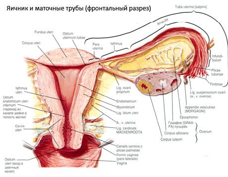
The paroophoron is a small structure that is also located in the mesentery of the fallopian tube, near the tubular end of the ovary. The paroophoron consists of several separate blind tubules.
Vesicular appendices (appendices vesiculosae), or stalked hydatids, look like bubbles that are attached to long stalks and contain a transparent liquid in their cavity. Vesicular appendages are located lateral to the ovary, slightly below the lateral part (infundibulum) of the fallopian tube.
 [
[ What do need to examine?