Medical expert of the article

New publications
Congenital aneurysm
Last reviewed: 29.06.2025

All iLive content is medically reviewed or fact checked to ensure as much factual accuracy as possible.
We have strict sourcing guidelines and only link to reputable media sites, academic research institutions and, whenever possible, medically peer reviewed studies. Note that the numbers in parentheses ([1], [2], etc.) are clickable links to these studies.
If you feel that any of our content is inaccurate, out-of-date, or otherwise questionable, please select it and press Ctrl + Enter.
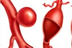
A pathologic weakening and subsequent localized bulging of the wall of an arterial vessel, ventricle of the heart, or interatrial septum that occurs due to a congenital defect or genetic disease is diagnosed as a congenital aneurysm.
Epidemiology
Syndromal aneurysms account for 10-15% of cases; thoracic aortic aneurysms and dissection occur in 80% of patients with Marfan syndrome.
According to studies, the prevalence of pulmonary artery aneurysms in congenital heart disease is about 6% of cases, and the prevalence of congenital left ventricular aneurysms in adults is no more than 0.7%. Atrial septal aneurysms are found in 1% of children and 1-2% of adults.
The incidence of aortic sinus aneurysm is estimated to be 0.09% of the general population, and it represents 3.5% of all congenital heart defects.
Cerebral aneurysms develop in approximately 50% of patients with congenital cerebral arteriovenous malformations, but they account for less than 5% of cases in children under 12 years of age. According to clinical statistics, 85% of all intracranial aneurysms occur in the vessels of the circle of Willis in the brain.
Causes of the congenital aneurysm
Noting the causes of congenital aneurysms, experts put genetically determined pathologies of connective tissue in the first place:
- Marfan syndrome;
- Ehlers-Danlos syndrome (vascular type);
- Loes-Dietz syndrome.
Arterial aneurysms form more easily in patients with williams syndrome (Williams-Boyren), which results from a deletion of the ELN gene, which encodes the tropoelastin protein, a precursor to elastin (an important structural element of the extracellular matrix of vascular wall tissues).
Non-syndromal hereditary (familial) thoracic aortic aneurysms are also defined, and mutations in some genes (MYH11, ACTA2, etc.) have already been identified.
Aneurysms of congenital origin can form in the presence of genetic enzyme abnormalities - different types mucopolysaccharidosis in children, which arise from mutations in genes that encode enzymes needed to metabolize glycosaminoglycans (mucopolysaccharides).
Cerebral aneurysms present at birth are not only associated with syndromal collagenoses. Such focal bulges of the cerebral vessel walls of the cerebral arterial circle, the circle of Willisia, are observed in:
- Arteriovenous malformations - a congenital vascular pathology, the essence of which is a violation of the architectonics of arterial and venous vessels, leading to changes in the normal blood flow;
- Autosomal dominant polycystic kidney disease.
Congenital aneurysm formation of the abdominal aorta or renal arteries can be neurofibromatosis type 1, a genetic disorder that results from a spontaneous mutation in nearly half of patients.
The causes of venous aneurysms (which are rare in clinical practice) are considered to be vascular malformations formed during intrauterine development (e.g., in Servel-Martorell syndrome), as well as venous angiodysplasia of the vein dilation type in Klippel-Trenaunay syndrome, which has been found to have a genetic basis.
Read also:
Risk factors
There is an increased risk of congenital cardiac aneurysm formation in the presence of congenital heart defects, and left ventricular wall bulging of the heart in the presence of congenital hypertrophy. [1]
A thoracic aortic aneurysm is more likely to form in the case of a congenital bicuspid aortic valve defect. But the threat of pulmonary aneurysm is much higher if there is open oval window in newborns, especially premature infants. [2]
Intracranial aneurysms form more frequently than others in children with congenital coarctation of the aorta, as well as with constitutional vasculopathies, particularly anomalies of the vessels of the circle of Willis (of which arterial hypoplasia is considered the most common). [3], [4]
In almost all cases, genetic risk factors and the presence of a family history of aneurysms, which suggests a predisposition to their formation, play a critical role.
Pathogenesis
The main pathogenesis of aneurysms of the main blood vessels in Marfan syndrome is a mutation in the FBN1 gene and impaired production of the extracellular matrix protein of the vascular wall fibrillin-1. This alters the structure and mechanical properties of the media, the middle sheath (tunica media) of arterial vessels. In addition to aneurysms of the ascending segment of the aorta, patients with this syndrome have aortic valve insufficiency.
The mechanism of aneurysm formation (including the splenic artery) due to the vascular type of Ehlers-Danlos syndrome is explained by certain gene mutations that lead to fragility of collagen types I and III - fibrous proteins of the intercellular substance of the vascular wall tissue. Such changes also weaken the vessel wall, and under the action of hemodynamic forces of blood flow in the weakest place or in the area of increased "hemodynamic stress" it gradually expands and bulges.
The pathogenesis of arterial aneurysms in Loes-Dietz syndrome associated with mutations in the genes for the transforming growth factor receptor (TGF-β), experts believe in the fragmentation and loss of elastin, the fibers of which provide reversible elasticity of the walls of large blood vessels. Damage of these fibers leads to a decrease in the contractility of the vessel wall. At the same time, proteoglycan molecules accumulate in the medial intercellular matrix, which reduces the stability of type I collagen, which maintains the strength of vascular walls.
In mucopolysaccharidosis, there is an accumulation of glycosaminoglycans, large linear polysaccharides (carbohydrates) that disrupt the arrangement of collagen fibrils, in the endothelial and medial lining cells of the vessel wall.
In addition to progressive renal cyst formation, PKD1/PKD2 mutations in polycystic kidney disease are thought to cause collagen defects, and patients with this inherited disease have aortic and cervico-head artery dissection and aneurysm formation - of the coronary arteries, interatrial septum, and intracranial vessels.
As histologic studies in recent years have shown, in cases of neurofibromatosis type 1, aneurysm formation of the abdominal aorta or renal arteries may be led by proliferation of Schwann cells of the peripheral nervous system, which spread to the sheaths of the vascular wall.
In cerebral arterial vascular defects, the mechanism of cerebral aneurysm formation is associated with disturbances in the distribution of blood flow: carotid (i.e., blood flow in the internal carotid arteries - arteria carotis interna) and basilar - in the main artery of the posterior part of the brain (arteria basilaris) and its branches.
Symptoms of the congenital aneurysm
According to their shape, there are such types of aneurysms as sac-shaped and spindle-shaped (fusiform), and according to their localization they are divided into types: cerebral (aneurysms of cerebral vessels), aneurysms of the thoracic or abdominal aorta, aneurysms of peripheral arteries and others.
Congenital aortic aneurysm
Aneurysms affecting any part of the aorta from the aortic valve to the iliac arteries. [5] Their symptoms are detailed in publications:
- Thoracic aortic aneurysm
- Aneurysm of the ascending aorta.
- Abdominal aortic aneurysm
- Aortic branch aneurysms
Congenital aortic sinus aneurysm
The aortic sinus or sinus of Valsalva is one of the anatomical dilations of the ascending aorta immediately above the aortic valve. Aortic sinus aneurysm is localized between the aortic valve ring and the region of the ascending aorta where the configuration of the vessel becomes tubular again (so-called sinotubular junction). Such an aneurysm is formed due to weakening of the elastic lamina of the vessel wall and forms as a blind diverticulum. An unruptured aortic sinus aneurysm is usually asymptomatic, but if it is large enough, it may manifest with cardiac arrhythmias, aortic regurgitation, atrial fibrillation and lead to acute coronary syndrome. [6], [7]
Congenital cerebral vascular aneurysm
A small aneurysm may not cause any symptoms, but the first signs of enlargement are sudden onset of severe headaches, nausea and vomiting. A large aneurysm may press on brain structures, which is manifested by pupil dilation and double vision, eyelid drooping, pain above and behind one eye, numbness on one side of the face, and seizures. [8], [9]
More information in the material - arterial aneurysms of cerebral vessels
Congenital cardiac aneurysm
All details in the publication - acute and chronic cardiac aneurysms: ventricular, septal, postinfarction, congenital
Congenital atrial septal aneurysm
An atrial septal aneurysm is a congenital malformation of the atrial septum, a localized sac-like deformity that usually arises in the oval fossa and often bulges into one or both atria during the cardiac cycle. [10]
The pathology may be associated with congenital heart defects, particularly open oval window, valve prolapse, and open ductus arteriosus, but in one-third of cases it manifests as an isolated anomaly.
Among the main symptoms are noted: shortness of breath, especially with physical exertion; fatigue; swelling of the lower extremities and abdomen; arrhythmia and palpitations; heart murmurs (when listening). [11]
Congenital venous aneurysms
These are rare malformations - vascular anomalies that can occur in most large veins; the most common localization is the lower extremities (more than 75% of cases), upper extremities (up to 10% of cases). Aneurysms of the internal jugular vein (most often spindle-shaped) account for 13% of cases, with two thirds of patients being children and adolescents. [12]
A superficial vein aneurysm manifests as a soft subcutaneous mass (usually painless) that enlarges with pushing, coughing, or crying. [13]
Complications and consequences
Aneurysm walls are pathologically altered vessel walls that have lost their normal biomechanical properties, and the extensibility of their bulging site is limited, so the main consequences and complications of any aneurysm are rupture.
All details in the publication - ruptured thoracic and abdominal aortic aneurysm
In this case, rupture of a congenital cerebral vascular aneurysm is accompanied by subarachnoid hemorrhage, resulting in a pediatric mortality rate of more than 10%.
Rupture of an aortic sinus aneurysm is a dangerous complication: rupture of the right and non-coronary sinuses usually leads to a connection between the aorta and the right atrium or right ventricular outflow tract, resulting in left-to-right discharge. This can cause right ventricular overload and right-sided heart failure.
A complication of a congenital aneurysm of the heart, particularly of the left ventricle or atrial septum, can be extrinsic thrombosis (due to blood stasis); the clot can break off, with the potential for peripheral arterial embolism and cryptogenic stroke. And when such an aneurysm ruptures, there is cardiac tamponade.
Diagnostics of the congenital aneurysm
To diagnose a congenital aneurysm, a comprehensive examination is necessary, the basis of which is instrumental diagnosis, which includes: ECG and echocardiography; ultrasound and computed tomography of the chest; ultrasound of the arteries of the internal organs of the abdominal cavity; coronary angiography; and aortography; contrast ventriculography; multispiral CT angiography and MR angiography; CT scan of the skull base; transcranial Dopplerography, etc.
Differential diagnosis
Differential diagnosis can be challenging. For example, ventricular septal aneurysm should be differentiated from large septal defects, ventricular hypertrophy, Fallot's tetrad and Eisenmenger's syndrome. And venous aneurysms should be differentiated from varicose veins and truncular venous malformations, hemangioma, laryngocele, and enteric cyst.
Who to contact?
Treatment of the congenital aneurysm
Treatment usually starts with a medicine that helps lower blood pressure or relax blood vessels to prevent the aneurysm from rupturing. Anticoagulants are also used - to prevent blood clots from forming.
Read:
In symptomatic congenital aneurysms, surgical treatment is performed using various techniques (including endovascular) depending on the localization and size of the aneurysm. Read more in the article - surgery for arterial aneurysms
Prevention
As preventive measures available today, experts point out: medical and genetic counseling, genetic analysis in pregnancy, as well as prenatal diagnosis of congenital diseases.
Forecast
Unfortunately, there cannot be a favorable prognosis for all cases of congenital aneurysms. For example, how long do patients with genetically determined syndromes with connective tissue pathology live? In general, the average survival rate for Marfan syndrome is about 70 years, for Ehlers-Danlos syndrome about 50 years, and for Loes-Dietz syndrome just over 35 years. And in most cases, death occurs from ruptured aneurysms, cerebral hemorrhage, or thoracic/abdominal aortic dissection.
Patients with ruptured aortic sinus aneurysms usually die within one year of diagnosis - due to congestive heart failure, according to clinicians.