Medical expert of the article

New publications
Moles in children
Last reviewed: 04.07.2025

All iLive content is medically reviewed or fact checked to ensure as much factual accuracy as possible.
We have strict sourcing guidelines and only link to reputable media sites, academic research institutions and, whenever possible, medically peer reviewed studies. Note that the numbers in parentheses ([1], [2], etc.) are clickable links to these studies.
If you feel that any of our content is inaccurate, out-of-date, or otherwise questionable, please select it and press Ctrl + Enter.
Many of us have heard that birthmarks can be dangerous. However, as well as that all birthmarks should be treated with special care. That is why birthmarks in children are not an unfounded reason for concern on the part of parents. After all, all mothers and fathers want to see their babies healthy and beautiful. If a birthmark is small and located somewhere on the arm, back or bottom of the child, it causes affection in parents. Another matter is significant spots of irregular shape, located on the face and other places not hidden by clothing. They are not only aesthetically unattractive, but can also conceal a hidden threat to the life of the child.
 [ 1 ]
[ 1 ]
Causes baby moles
Moles are unusual neoplasms on human skin. The mystery of these pigment spots is that they can appear at any age, starting from the moment of birth of the child. True, the presence of moles (nevi) in newborns is a rather rare phenomenon, which occurs in one baby out of a hundred. Nevertheless, the fact remains that the child can already be born with a mark, which is called a birthmark. A birthmark can have a more or less saturated brown or red color and be of different sizes.
Usually, moles begin to appear on children's skin starting from the age of six months, but in most cases this process starts at 2-3 years. By the age of four, most children can see about 10 moles of different sizes on their skin. Then, for some time, the increase in the number of pigment spots does not occur or slows down. The next peak in the growth of the number of nevi falls on adolescence, when the appearance of moles is associated with hormonal changes in the body.
In principle, the appearance of moles in humans is a natural process. This is due to the presence of special cells in human skin - melanocytes, which in some cases cause various changes in the pigmentation of the skin.
The reasons for the appearance of moles in a child can be either hereditary or the result of internal (changes in hormonal levels during puberty) and external (influence of sunlight) influences. If the child has numerous birthmarks in his family, then, most likely, he will have many moles. Moreover, they appear mainly in the same places as in relatives, which, by the way, is the reason for the name of such neoplasms.
During adolescence, hormonal surges can cause an increase in the production of melanin, a substance responsible for skin pigmentation. During puberty, nevi can both actively appear and disappear. Moreover, such behavior of moles does not indicate pathological processes in the body or directly on the skin. This is a normal, natural reaction.
There is also a theory that changes in skin pigmentation can be caused by traumatic effects on the skin, such as insect bites, or the influence of viral infections that trigger the process of grouping and exiting melanocytes to the surface. There are moles that are almost invisible on the skin. A child can accidentally scratch it, and it will change color to a darker color.
The effect of ultraviolet radiation on the skin can also provoke an increase in the number of nevi, as well as a change in their appearance (color, size, shape). Moreover, this happens at any age, both in childhood and adolescence, and even in adulthood. It is the effect of ultraviolet radiation that can subsequently trigger pathological processes of modification and degeneration of moles.
Some studies show that the probability of birthmark formation in newborns is higher if the child is premature or has very light skin. Light-skinned children often have more birthmarks than dark-skinned children. There is a dependence of the number of nevi on the sex of the child. As a rule, girls are more likely to develop birthmarks.
[ 2 ]
Symptoms baby moles
As mentioned above, moles can have different shapes, sizes and colors. The color range of moles in children ranges from dark beige, almost invisible on the skin, to deep red and even black. Common safe moles in children have a regular round shape with smooth edges, brown color and small size up to 1.5 mm. They can be completely flat or slightly protrude above the surface of the baby's skin. Parents should not worry about such neoplasms.
Moles of medium (up to 10 mm) and large (more than 10 mm) sizes have a greater chance of being damaged and scratched, and accordingly, the probability of degenerating into a malignant neoplasm is higher. A good indicator is the presence of hair on the mole itself, regardless of its size. Such moles are not prone to degeneration if you do not pull out the hairs on them.
In addition to this division, in medical practice there is a division of moles by appearance and method of formation into common and vascular nevi. Common moles are smooth neoplasms of light pink or brown color. Sometimes their color is darker, but this should not frighten parents.
A black smooth mole on a child is more the norm than an abnormality. A rich dark color in this case is not an indicator of its danger to the baby's life. Another thing is if the mole changes color to a more or less rich shade, there are a lot of such moles, or if there is one black mole, but it is large (more than 1.5 cm). This is already a reason to consult a dermatologist.
A red birthmark on a child indicates its vascular origin. Vascular birthmarks are so named because they consist of a large cluster of blood vessels, and accordingly have a red color. They can have different shapes, and their color varies from light pink to deep red.
Vascular birthmarks in children come in different types and shapes:
- Hemangioma
- "Stork bite" - markings on newborns are a rich red-orange color
- "Port wine stains" - brownish-red or burgundy growths (flame nevus)
Hemangioma is a benign formation on the skin, despite its unaesthetic appearance. Their appearance may not be noticed immediately. This may happen 2-3 weeks after the birth of the baby or even after a year. Such a mark can have different sizes and locations. Its peculiarity is the ability to grow. Even if such a birthmark grows very quickly in a child, it does not pose a danger to life, except for discomfort from an aesthetic point of view. Usually, by the age of one and a half years, hemangiomas become much lighter, and by the age of 10 they disappear completely.
There are 2 types of hemangioma: "strawberry" and "cavernous". "Strawberry" mole is soft to the touch, has a convex structure and a color similar to the berry of the same name. Such moles most often appear on the face of a child, as well as on the head, back of the head and neck, but their appearance in other places, including even internal organs, is not excluded.
"Cavernous" hemangioma looks a little different. It has a purple, deep burgundy or bluish-gray hue, a denser structure, going deep into the layers of the skin. Often, this is an irregularly shaped spot, consisting of one or more foci close to each other. It can appear on different parts of the body.
The biggest frustration for parents is caused by birthmarks on the child's face and head. But you just need to be patient, because such growths disappear on their own. Usually, they are not treated. It is simply necessary to take all measures to ensure that the baby does not damage such a birthmark, does not scratch it. After all, the main reason for the transformation of a birthmark into a life-threatening neoplasm is its injury. And the larger the birthmark in size and the more it stands out above the surface of the skin, the higher the probability of its damage.
The most common mark on the face and back of the baby's head is a yellow or cream-red birthmark, jokingly called a "stork bite" (or "angel kiss"). This can be a single large pink or cream-colored spot or a cluster of several spots. These marks usually disappear by the age of one year, but there are cases where they remain for a longer period.
The situation is more complicated with "port wine stains" - flat smooth neoplasms of a red-burgundy color. Such birthmarks in children also tend to increase in size as the child grows, but do not disappear with age. They cannot be removed. You can only try to make them less noticeable with the help of home remedies for lightening skin spots or professional cosmetics. In some cases, a course of infrared or laser therapy can be recommended.
It is worth noting that some parents mistakenly believe that such a spot can be hidden by a tan and allow children to stay in the sun for a long time. Such a careless attitude can only lead to a change in the color of the spot to a more saturated one, but will not hide the defect in any way. In addition, increased exposure to ultraviolet rays of the sun can lead to the degeneration of the mole.
A hanging mole in a child has a special place among birthmarks. It can be located on the baby's neck or under the arms. It can appear at any age. Such a mole looks like a small piece of hanging skin of a natural or darker color. The whole danger of a hanging mole is that it cannot be torn off or injured, while it can become the object of close attention of your baby. Removing such a mole on your own is also not worth it if you care about your child's health. The most correct solution would be to consult a dermatologist for an examination and consultation, as well as close monitoring of the behavior of the hanging mole: for changes in the color and size of the nevus.
Symptoms of degeneration of moles
In general, if a mole is not injured during life and does not undergo any visible changes, it exists on the body of its owner for a long time without causing harm to his health. This is typical mainly for small moles up to 6 mm in diameter. A dangerous mole in a child is one that is larger than 6 mm. It is dangerous not in itself, but because the risk of injury to such neoplasms is higher than to small spots.
The same applies to a convex birthmark in a child. A child, having felt an unusual lump on his body, will show special attention to it. He may constantly touch it, try to tear it off. The risk of injury to such birthmarks is very high, so it is necessary to carefully monitor not only the behavior of the birthmark, but also the child's actions in relation to it.
A large birthmark on a child, no matter when it appears and no matter how it looks, is certainly a reason to show the baby to a dermatologist. The doctor will be able to assess the likelihood of the birthmark becoming malignant and will definitely give advice on caring for nevi.
The pathogenesis of the transformation of harmless birthmarks and moles in children into dangerous malignant neoplasms has not yet been fully studied by doctors, however, the causes of these changes have been reliably determined. These are trauma to the surface of the birthmark, unsuccessful attempts to remove birthmarks using questionable methods and means, as well as prolonged exposure to the sun without the necessary protection.
The consequences and complications of the influence of these causes can be the most tragic. Injury to a mole can lead to ulcers and bleeding from the nevus, which are very difficult to stop. At the site of the lesion, in this case in the area of the mole, a malignant tumor (melanoma, or skin cancer) can develop, which has a very rapid development with multiple metastases in all parts of the body. At the same time, early symptoms of melanoma development detected in time guarantee a 95% probability of successful treatment. If the disease is neglected, this probability drops to 20%, the remaining 80% of cases lead to the death of the patient.
Any birthmarks on the baby's body require attention from parents. Periodic examination of birthmarks will allow you to notice the first signs of a birthmark changing and turning into a malignant tumor. Such signs include:
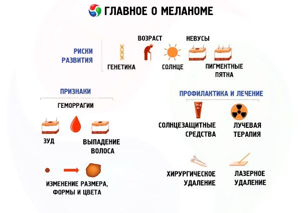
- Asymmetry of the neoplasm (asymmetry). Ideally, a mole is a circle or oval, the two halves of which are symmetrical (similar) to each other. If one side of the mole grows more than the other, this is already a reason to examine it.
- Uneven borders of a nevus (border irregularity). A normal healthy mole always has even edges. If the borders of a nevus become blurred, with jagged edges, this is already one of the signs of melanoma development.
- Color change. A uniform color of a pigment spot is considered normal. Inclusions of any color on the uniformly colored surface of a nevus become noticeable to the eye. Any strange birthmark in a child with an unusual color or shape should alert caring parents.
- Mole diameter (diameter). If the mole is no more than 6 mm in diameter, then there is no need to see a specialist. Regular periodic monitoring is sufficient. Moles with a larger diameter are best shown to a dermatologist immediately to assess its development and growth.
- Evolving behavior. As a rule, a mole does not undergo any significant changes during a person's life. However, if any of the above characteristics or several of them at once begin to change, it is better to immediately show the child to a dermatologist or oncologist to prevent sad consequences. The appearance of a large number of similar ones around the nevus should also be a warning sign.
This method of examining a mole for benignity and safety is commonly called the ABCDE method.
Where does it hurt?
Complications and consequences
Not all changes in moles in children and the area around them indicate the possible onset of skin cancer. For example, if a child's mole has grown, this may be both the beginning of a pathological process and a natural physiological manifestation. After all, birthmarks grow with children. In this case, it is worth seeing a dermatologist, but you should not "wind yourself up" in advance. If a mole has noticeably increased in size in a short period of time (within a month), then you should definitely not postpone a visit to the doctor.
A white spot around a mole in a child is not dangerous at all. Such a mole with contrasting pigmentation is called a nevus of Sutton. It can be a consequence of sunburn on the skin, when a spot with intense pigmentation forms inside, and its halo has no pigmentation at all. Such nevi disappear after a few years on their own, leaving no traces.
If a child's mole itches, it may be a sign of dry skin or a lack of vitamins in the body. However, it is risky to ignore this symptom, because it may also indicate the beginning of the degeneration of the mole, especially if other changes are superimposed.
Doctors also have an ambiguous attitude towards the appearance of a rough mole in a child. On the one hand, intradermal moles in infants, which are benign neoplasms, have a rough structure similar to a blackberry. On the other hand, moles should have a fairly smooth surface, and the appearance of roughness should be a warning sign. Moreover, such a mole can crack and bleed in the future, an infection can get into it and lead to inflammation of the skin in this area and other dangerous consequences. In any case, the child should be shown to a dermatologist, who will tell you what to do next and whether you should worry about the structure of the nevus.
If a child's mole hurts, it is often a consequence of its injury. It is necessary to examine the nevus for damage and, if any, treat the wound with a disinfectant solution. Do not wait until the pain goes away, even if it is insignificant. It is better to immediately consult a doctor and thereby prevent negative consequences. The same should be done if there is no external damage, but the mole continues to hurt. This may indicate the onset of pathological changes in it.
The red color of a mole in children always alarms parents. But some types of moles (angiomas) initially have this color and do not pose a danger if they are not injured. If a mole in a child turns red with a change in color to a more intense color or simply changes color from brown to red, this already indicates the presence of an inflammatory process in it associated with trauma or exposure to ultraviolet rays. In this case, a visit to a dermatologist and even an oncologist should be immediate in order to prevent the possible development of a tumor in time.
Diagnostics baby moles
Having noticed a strange birthmark on the child's body or having discovered a suspicious change in it, parents immediately have a question: where to go for advice and where can I check the children's birthmarks for benignity? Dermatologists are involved in the diagnosis and prognosis of the behavior of birthmarks, which means that it is necessary to contact them first. If the dermatologist suspects the development of malignant processes in the birthmark, he can refer the little patient for examination to a dermato-oncologist, or, if there is none, to a regular oncologist.
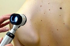
The most popular method of instrumental diagnostics of moles is dermatoscopy. In the past, a microscope was used for this purpose. In modern medical research, preference is given to a special device called a dermascope, which allows, with multiple magnification, to examine the slightest changes in the structure of a mole in children and adults.
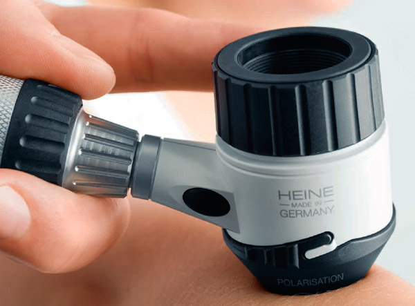
After the examination, the patient receives a photograph of his mole with its full description. The results of the dermoscopic examination are subsequently used in the differential diagnosis of nevi and their changes.
The second most popular, but even more accurate method of examining moles is computer diagnostics of pigment spots, or scientifically digital dermatoscopy. It allows you to get an image of a mole in tenfold, and even hundredfold magnification, to determine with great accuracy all the parameters of the nevus and its borders.
The high image accuracy allows you to see the smallest details, such as melanin spots, the slightest color changes in spots, and blood vessels on the skin surface. Some digital video dermascopes are able to detect the presence of altered cells that indicate the development of skin cancer.
A schematic representation of the location of moles on the patient's body is saved and entered into a database, which allows for comparative analysis during subsequent visits to the doctor.
And yet, both of these methods can only suggest the presence of malignant processes in a mole, but only histological tests (biopsy), which are carried out after the removal of a suspicious neoplasm, can show exactly whether oncology is present in this case or not. For histological examination, the cells of the excised mole that survived after the operation are taken.
What do need to examine?
How to examine?
Who to contact?
Treatment baby moles
A birthmark is not a pimple that can be cauterized with an alcohol-containing product and it will disappear. Treatment of birthmarks most often consists of surgical or laser removal. Birthmarks in children are removed mainly by the second method, as it is less painful and leaves virtually no unaesthetic scars at the site of the neoplasm. In addition, laser therapy helps prevent the development of tumor metastases.
However, such operations are most often performed in the case of transformation of a benign mark into a malignant tumor. The second indication for surgical removal of moles in children may be such a size, shape and location of the mole that greatly increase the likelihood of its injury, including by clothing (on the neck in the collar area, under the arms, on the palms and feet, etc.).
Conservative treatment of moles in children is carried out very rarely and according to the doctor's instructions. In this case, the age and weight of the little patient are taken into account. The same methods and means are used as for the treatment of adults. But sometimes the treatment can only aggravate the process, so it is necessary to weigh all the pros and cons a hundred times before deciding on such a step.
Treatment without mole removal is usually prescribed for red moles, and only if they change shape, start to grow, or there is a risk of frequent injury to the nevus. For the treatment of small angiomas, the doctor may prescribe cryodestruction (low-temperature exposure for several seconds), which helps to positively solve the problem in 96% of cases.
Simple angiomas of large sizes can be subjected to hormonal therapy with drugs. For this purpose, "Prednisolone" is used at a rate of 4-6 mg per 1 kg of the child's body weight, starting from 2 months of age. A two-time intake is recommended: a third of the daily dose at 6 am and two thirds at 9 pm. The course of treatment is 4 weeks with the drug taken every other day.
Intradermal modified moles in children on closed parts of the body are most often subject to laser removal. If such marks are observed on the face, sclerotherapy may be used, when drugs are injected under the skin that lead to the death of mole cells under the skin without damaging the outer layers. Such drugs include: quinine-urethane, hydrocortisone, 70% alcohol, 10% sodium chloride solution. The course of treatment is long, consisting of 10-15 injections, which are made once every 2 weeks or once a month.
In case of deep and extensive hemangiomas, angiography is performed before the start of treatment to determine the nature of the blood supply to the mole. Then, according to indications, hydrogel embolization is performed, which reduces the blood supply to the tumor and causes its reduction. The treatment ends with cryodestruction without subsequent removal of the mole. It itself subsequently dissolves, leaving behind an unaesthetic mark, which is removed by plastic surgery at an older age.
Folk treatment of moles
It cannot be said that folk remedies are useless or even dangerous for treating moles. But when it comes to a child, before using such remedies, it is necessary to examine the baby with a dermatologist to exclude the possibility of the mole degenerating into a cancerous tumor. After all, folk remedies can "dry up" a mole or lighten it, but they are not suitable for fighting skin cancer.
In the presence of malignant processes, such treatment can only worsen the situation, and you will lose precious time. Any treatment of moles in children should be carried out only after a detailed consultation with a doctor regarding the use of various methods and means, including traditional medicine.
For those who decide to try old grandma's or more modern folk methods of combating moles, we provide some recipes:
- Hydrogen peroxide applied to a mole 3 times a day will help get rid of it forever within a week or make the pigment spot less noticeable.
- Garlic and lemon juice. Apply one or the other juice to the moles several times a day for a week until they disappear.
- You can lubricate the mole with a mixture of crushed chalk and hemp oil. This will supposedly help make the mole less noticeable.
- Sometimes, onion juice is used to remove a mole, which is applied to the nevus several times a day.
- You can lubricate the mole with honey until it disappears. This delicious recipe will not cause any harm and your baby will even like it if he is not allergic to bee products.
- A gruel made from black radish helps to get rid of a mole if you apply it to the spot 4 times a day, covering it with a bandage.
- You can also do the same with a pulp of unripe apple mixed in equal proportions with honey.
- Applying a gruel of raw potatoes to a mole helps reduce the pigmentation of the mole to a barely noticeable spot.
In folk medicine, herbal treatment is also used to remove and lighten moles in children and adults.
- The juice of the milkweed herb will help get rid of a mole if you lubricate the nevus with it for a long time.
- A good effect is achieved by applying crushed dandelion root to the mole. This should be done every day, leaving the composition for several hours, securing the place with a plaster.
- Celandine juice acts similarly to hydrogen peroxide when used three times a day.
- Lightening a mole can be achieved by rubbing it with parsley juice several times a day.
As we can see, folk medicine uses both gentle and rather aggressive methods to combat moles. And it is up to you, the parents, to decide whether to test them on your child or not.
More information of the treatment
Prevention
It is impossible to protect a child from the appearance of moles, since heredity plays a decisive role in their occurrence. Nevertheless, some precautions should be taken, because the prognosis for the development of the neoplasm will depend on this: whether it will remain harmless throughout the baby's life or turn into a severe "headache" for parents and the child.
This primarily concerns the child's exposure to the sun, especially in spring and summer. The head of a small child must be covered with a Panama hat or scarf, the rest of the skin - with light "breathable" clothing. This measure will prevent the appearance of new nevi, as well as the degeneration of existing ones.
Parts of the body not protected by clothing, especially when on the beach, should be lubricated with sunscreen. Today, there are a great many sunscreens for children on sale in the form of creams, emulsions, sprays and body lotions with varying degrees of protection from the negative effects of UV rays.
Keep an eye on your child, and if you notice increased attention to the mole on his part, try to explain that touching and scratching the mole is not allowed. If the child is small, you can try to hide the mole under clothing, but under no circumstances under a bandage. Teenagers can try to get rid of the hated mole on their own. Explain to them that this should not be done without consulting a doctor.
Moles, which are more susceptible to injury than others, are best removed immediately, without waiting for a sad event. This will prevent damage to the nevus, which can subsequently cause the development of melanoma.
Systematic examination of moles in a child, which helps to identify pathological changes at an early stage of the disease, makes the prognosis for the treatment of dangerous nevi generally positive. In 95% of cases of melanoma development at the site of moles, a complete cure occurs.
Moles and birthmarks are areas of reduced skin immunity, which means they are more susceptible to negative degeneration than others. In addition, they often arouse increased interest in young researchers who can accidentally damage a nevus, especially if it stands out significantly above the skin surface, as in the case of hanging moles, or has a bright color in the case of angiomas. This is why moles in children should always remain the object of close attention from their parents, because untimely or incorrect treatment of such neoplasms can cost the child his life.