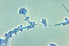
The causative agent of sporotrichosis (Sporothrix schenckii)
Last reviewed: 23.04.2024

All iLive content is medically reviewed or fact checked to ensure as much factual accuracy as possible.
We have strict sourcing guidelines and only link to reputable media sites, academic research institutions and, whenever possible, medically peer reviewed studies. Note that the numbers in parentheses ([1], [2], etc.) are clickable links to these studies.
If you feel that any of our content is inaccurate, out-of-date, or otherwise questionable, please select it and press Ctrl + Enter.

Sporothrix schenckii causes sporotrichosis (Schenck's disease) - a chronic disease with local damage to the skin, subcutaneous tissue and lymph nodes; possible defeat of internal organs. The causative agent was first described by Schenck in 1898.
Morphology and physiology
Sporothrix schenckii is a dimorphic fungus. In the patient's body, it grows in the yeast (tissue) form, forming cigar-shaped, oval cells 2-10 microns in diameter. Asteroid bodies (10-211 μm) are also found. Asteroid bodies are formed by yeast-like cells and are surrounded by ray-like filaments and rays. On the nutrient medium (glucose agar Saburo, 18-30 ° C), the fungus forms folded white or dark colonies consisting of a thin septate mycelium (mycelial form) with cones of oval conidia in the form of daisy flowers. There are also sedentary (on hyphae) conidia of a darker color. Conidia (spores) are connected by hyphae-hairs, hence the name Sporothrix.
Pathogenesis and symptoms of sporotrichosis
At the site of penetration of S. Schenckii through the damaged skin formed an ulcer of irregular shape, nodules and abscesses. Fungus spreads lymphogenously. In the course of the proximal lymphatic tract, nodules are formed, followed by ulceration. The most common form of the disease is lymphatic (lymphatic) sporotrichyosis. The affected areas are densified and painless. Nodular skin lesions can also occur with mycobacteriosis caused by opportunistic mycobacteria (M. Marinum, etc.).
Sometimes there is dissemination of the pathogen with the development of visceral sporotrichyosis: the lungs, the bone system, the abdominal organs and the brain are affected . Perhaps the development of primary pulmonary sporotrichyosis. When the disease appears, antibodies develop HRT. Fungi are destroyed by neutrophils and macrophages.
Epidemiology of sporotrichosis
In the mycelial form, S. Schenckii lives in soil and rotting plant material; it is found in wood, water and air. Distributed in the tropics and subtropics. People with agricultural work are more often ill. The causative agent gets into areas of skin microdamages by contact (a disease of working with roses). Possible penetration of the fungus through intact skin or its entry into the lungs by an aerogenic mechanism.
Microbiological diagnosis of sporotrichosis
Examine the allocation of ulcers, microabscesses, skin, punctate lymph nodes and tissues. Preparations are stained with hematoxylin and eosin, according to Romanovsky-Giemsa, Gram-Weigert, acridine orange. When microscopic examination of a smear or a biopsy from a lesion focus, yeast-like cells and asteroid bodies of the fungus are identified. A pure fungus culture in the form of a mycelial phase is isolated by culturing on nutrient media at 22-25 ° C for 7-10 days (at 37 ° C the yeast form of the fungus develops). When interteasticular introduction of guinea pigs to a grown mycelium, it turns into a yeast form. In the blood serum of patients, antibodies in RA, RP, ELISA, etc. Are sometimes detected. An allergic test is placed with the allergen sporotrichin.


 [
[