Medical expert of the article
New publications
Vascular impingement in the cervical region
Last reviewed: 29.06.2025

All iLive content is medically reviewed or fact checked to ensure as much factual accuracy as possible.
We have strict sourcing guidelines and only link to reputable media sites, academic research institutions and, whenever possible, medically peer reviewed studies. Note that the numbers in parentheses ([1], [2], etc.) are clickable links to these studies.
If you feel that any of our content is inaccurate, out-of-date, or otherwise questionable, please select it and press Ctrl + Enter.

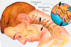
Cervical spine conditions can cause problems with pinched nerves, but there can also be pinched blood vessels in the cervical spine that interfere with blood flow to the brain.
Causes of the vascular impingement in the cervical region
The cervical spine contains such vessels as: the right and left vertebral arteries; the common carotid or carotid artery (which is divided into the right and left carotid arteries, and those, in turn, into the internal and external carotid arteries). The cervical part of the internal carotid arteries (a.carotis interna), through which blood flows to the brain, passes over the palatine tonsil - along the transverse processes of the cervical vertebrae: C3, C2 and C1. The external and internal jugular veins (with branches) also run in the cervical region.
One of the most important blood vessels of the neck is the vertebral arteries (a.vertebralis), which branch from the subclavian arteries at the base of the neck and pass through the openings of the transverse processes of the cervical vertebrae C6-C1.
The main causes leading to pinched blood vessels running in the cervical region include:
- Instability of the cervical spine due to disorders of its ligamentous apparatus, in particular, dislocation of tendons attaching muscles to the cervical vertebrae;
- Spondylolisthesis - cervical vertebrae displacement; [1]
- Cervical osteochondrosis with the formation of osteophytes (bone growths);
- Degenerative changes in the cervical spine - cervical spondylosis; [2]
- Deforming cervical spondyloarthrosis (with development of hypertrophic changes of intervertebral joints);
- Protrusion and herniated discs; [3]
- Cervical scoliosis. [4]
Cervical spine injuries may involve pinching of the cervical anterior spinal (spinal) artery (a. Spinalis anterior), which originates from the two vertebral arteries at the level of the greater occipital foramen and runs to the C4 cervical vertebra.
After a so-called whiplash injury to the neck, there may be increased mobility of the craniocervical junction or transition, which consists of the occipital bone of the skull base and the joints of the first two vertebrae of the neck (C1 and C2). As a result of the weakening of the ligaments that hold the head together - craniocervical instability - the internal jugular vein (v. Jugularis interna), which runs in front of the upper cervical vertebrae, is compressed. [5]
In rare cases, jugular vein compression may be caused by abnormal elongation (hypertrophy) of the styloid processus (processus styloideus) coming from the lower part of the temporal bone or calcification of the descending stylo-lingual ligament (ligamentum stylohyoideum).
The same cause, i.e. Excessive pressure of these structures and compression of the stylopharyngeus muscle (m. Stylopharyngeus) under the lower jaw may also be associated with impingement of the nearby internal carotid artery. In addition, in people with osteochondrosis of the cervical vertebrae, the carotid artery may be compressed by a spasmed anterior staircase muscle (m. Scalenus anterior), which flexes and rotates the neck.
Risk factors
Factors that increase the risk of pinched blood vessels in the cervical spine include: forced prolonged sitting (most often associated with professional activities) and sedentary lifestyle; trauma to the cervical spine; anomalies of the cervical spine or craniocervical junction; violation of lordosis of the cervical spine;presence of a cyst localized in the cervical spine; anterior ladder muscle syndrome; enlargement of lymph nodes - cervical and supraclavicular; osteoporosis; genetically determined connective tissue diseases; ossification of tendons and ligaments around vertebrae - diffuse idiopathic skeletal hyperostosis.
Pathogenesis
In explaining the pathogenesis of vascular impingement in the cervical region, it should be noted that the path of vertebral arteries in this segment of the spinal column passes in the bony canal, which is formed by the foramen transversarium of the cervical vertebrae. This is the only section of the spine that has openings in the vertebral bone for the passage of blood vessels. In addition to the vertebral artery and veins, sympathetic nerves pass through these openings.
Arteries and veins pass so close to bony structures that any damage to vertebral joints or their ligamentous apparatus, protrusion into the lumen of the foramen transversarium of the intervertebral disc (which may undergo ossification) or bony outgrowth (marginal osteophyte) can lead to impingement (compression, squeezing) of vessels with a decrease in their diameter and reduced blood flow rate.
For example, osteophytes of the hook-shaped processus (processus uncinatus) of a vertebra resulting from osteoarthritis of the Luschka joints (uncovertebral joints - synovial articulations between the bodies of cervical vertebrae C3-C7) may compress the vertebral artery when it passes through the opening of the transverse processes of the cervical vertebrae.That is, the mechanism of vessel impingement is due to stenosis (narrowing) of the transverse processus.
Symptoms of the vascular impingement in the cervical region
Arterial blood flow due to pinching of vertebral arteries is disturbed with deterioration of blood flow to the cerebellum, activating cerebral cortex reticular formation of the brainstem, inner ear. And the clinical picture of vessel pinching by osteophytes in cervical osteochondrosis or herniated disc bulge includes such symptoms as: pulsating headache (which becomes stronger when turning and bending the neck, as well as with any physical exertion); dizziness; noise in the head and ears; deterioration of vision with its "blurring", the appearance of "flies" and darkening in the eyes; impaired coordination of movements and balance or ataxia with subsequent weakness of the limbs; attacks of nausea and short-term loss of consciousness with sudden movements of the head.
When the common carotid artery is compressed below the carotid sinus (the point of dilation of the internal carotid artery at the level of the upper edge of the thyroid cartilage of the larynx), there is an increase in heart rate and blood pressure.
Signs of internal carotid artery impingement include numbness or weakness in a part of the body or on one side of the body; problems with speech, vision, memory, and thinking; and inability to concentrate.
Jugular vein compression is most commonly seen in the upper neck and can cause neck discomfort and stiffness, headaches, head noise, tinnitus or ringing in the ears, hearing problems, double vision, insomnia, and even transient memory loss.
Complications and consequences
The vertebral arteries supply blood to the brain stem, the occipital lobes and the cerebellum. The consequence of their impingement is vertebrogenic vertebral artery syndrome (Barré-Lieu syndrome), i.e. Vertebral artery compression syndrome. [6], [7]
Due to compression at the level of a.vertebralis and a.basillaris, blood flow in the vertebral-basilar system (cerebral arterial circulation circle) is weakened and vertebrobasilar insufficiency (Hunter-Bow syndrome) develops. [8]
Blockage of the cervical arteries can be complicated by vertebrogenic transient ischemic attacks, as well as acute disruption of blood supply to the brain and damage to its tissues - ischemic stroke. [9]
Impingement of the anterior spinal artery, which supplies blood to the upper spinal cord, leads to impaired spinal circulation, and arterial insufficiency is fraught with the development of ischemic spinal cord infarction. [10]
Diagnostics of the vascular impingement in the cervical region
Only instrumental diagnostics - x-ray of the cervical spine - can assess the condition of spinal structures; ultrasound Doppler vascular imaging, CT and MR angiography are used to examine vessels. Brain structures are visualized using magnetic resonance imaging.
Differential diagnosis
A differential diagnosis is made with peripheral vascular diseases (for example, narrowing of the lumen or stenosis of the carotid artery associated with atherosclerosis), pinched nerve in the cervical region (cervical radiculopathy), spinal cord compression.
Treatment of the vascular impingement in the cervical region
Comprehensive treatment of canal stenosis formed by the openings of the transverse processes of the cervical vertebrae depends on its cause and severity of the condition and includes:
- Drug treatment (including epidural injections of corticosteroids);
- Physical therapy;
- LFC;
- Therapeutic neck massage;
- Acupuncture.
Surgical intervention may be required. For example, in craniocervical instability, surgical fusion (spondylosis) - permanent immobilization of the joints of the C1-C2 vertebrae - is effective. Also possible prolotherapy - tightening the ligaments that hold the head, using special injections. And in case of styloid hyoid syndrome with compression of the jugular vein or carotid arteries, surgical intervention in the form of styloidectomy may be performed.
Prevention
To prevent pinching of the vessels passing in the cervical region, it is necessary to regularly perform exercises to strengthen the neck muscles, stabilize the vertebrae and train correct posture, as well as to ensure the correct position of the neck during sleep (with the help of an orthopedic pillow).
And should be timely treated leading to vascular congestion diseases.
Forecast
Given the possible complications of vascular impingement, the prognosis of its outcome, unfortunately, cannot be favorable for all patients.

