Medical expert of the article

New publications
Anterior ladder muscle syndrome
Last reviewed: 04.07.2025

All iLive content is medically reviewed or fact checked to ensure as much factual accuracy as possible.
We have strict sourcing guidelines and only link to reputable media sites, academic research institutions and, whenever possible, medically peer reviewed studies. Note that the numbers in parentheses ([1], [2], etc.) are clickable links to these studies.
If you feel that any of our content is inaccurate, out-of-date, or otherwise questionable, please select it and press Ctrl + Enter.
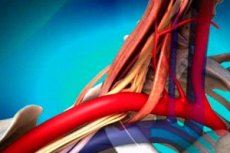
Anterior scalene syndrome (also called Naffziger syndrome, named after the author who first described this disease – HC Naffziger, 1937) is one of the most common variants of pathology in the collective category of syndromes observed in the upper sternal entrance.
Causes anterior ladder muscle syndrome
The cause of the syndrome is a reflex muscle spasm, which occurs due to irritation of the roots due to the development of cervical osteochondrosis. The anterior scalene muscle is located in the space between the transverse ends of the 3rd-6th cervical vertebrae, as well as the 1st rib. The lower region of the brachial plexus is also located in this place, together with the artery located under the collarbone - they are compressed due to the narrowing of the muscle.
 [ 4 ]
[ 4 ]
Pathogenesis
In Naffziger syndrome, the patient experiences compaction, spasm, or thickening of the indicated muscle, and in addition, secondary compression of the bundle of vascular-nerve endings (the vein and artery located under the collarbone, and along with them the bundle inside the brachial plexus, which is formed from roots of the C8-T1 type) in the area of the interscalene space (between the 1st rib and the spasmodic muscle).
Symptoms anterior ladder muscle syndrome
This syndrome is characterized by the following symptoms: pain in the neck area, which goes down the arm from the elbow, and in addition, painful sensations with tension in the arm. The pain syndrome becomes stronger at night, as well as in the case of a deep breath and an attempt to tilt the head to the healthy side. In some cases, the pain can go to the shoulder girdle, armpits and sternum. Carpal weakness may be felt (mainly in the 4-5 fingers), and sometimes vasomotor disorders in the hand are observed. Along with this, tingling with numbness may be felt in the hand, especially in the forearm, as well as on the ulnar carpal side.
As a result of compression of the artery due to spasm of the anterior scalene muscle, the supraclavicular fossa begins to swell, and in addition, the amplitude of arterial fluctuations and the level of blood pressure decrease (in the case of turning the head in the direction opposite to the compressed muscle). Along with this, the development of paresthesia in the upper limb, as well as headaches, is possible.
Blueness or pallor may also appear, as well as swelling in the area of the hand, and in addition, the skin temperature may decrease. Roughening of the skin, development of osteoporosis of the carpal bones, and development of brittle nails are possible. When palpating the compacted muscle, the patient feels pain.
Complications and consequences
In case of increasing manifestations of the syndrome, some complications may develop: hypotrophy of the wrist muscles, accompanied by symptoms of poor blood circulation in the limb. This condition is similar to Raynaud's disease (decreased pulse inside the radial artery, swelling of the wrist, occurrence of hypertensive crises caused by cold, etc.).
Diagnostics anterior ladder muscle syndrome
The diagnostic process is based on the clinical picture of the disease: as a result of palpation, one-sided swelling and thickening are found on the patient’s neck (to the right or to the left, depending on which muscle is being compressed) – this area is also painful.
To clarify the diagnosis, the so-called Edson test is performed: for this, the patient's arm is pulled back, and then he must throw his head back. As a result, the compression of the spasmodic muscle in relation to the subclavian artery increases. If the test is positive, the pain will increase and the arm will go numb. In this case, the pulsation in the area of the radial artery will weaken or disappear altogether.
During the diagnostic process, such instrumental procedures as rheovasography, oscillography, and in addition volumetric sphygmography are performed.
Differential diagnosis
During the diagnostic process, it is extremely important to distinguish in time the reflex contraction of the muscle inherent in the above-mentioned pathology from Pancoast syndrome, which has very similar symptoms and develops against the background of a tumor of the pulmonary apex.
Who to contact?
Treatment anterior ladder muscle syndrome
The main goal of the treatment course is to eliminate discomfort (numbness and pain), and in addition to this, to restore the natural healthy state of the vessels and muscles along with the motor function of the upper limb. At the initial stage, treatment is carried out using conservative methods.
During the treatment, various medications are used - a novocaine blockade of the spasmodic muscle is performed (hydrocortisone may be administered as an auxiliary agent). Diprospan may also be administered to the site of nerve compression. Along with this, anti-inflammatory drugs (salicylates with brufen), painkillers, and vasodilating drugs (such as no-shpa, complamin, and nikoshpan) are prescribed.
Complex conservative treatment also includes the use of vitamins from the B category.
Physiotherapeutic procedures include massage of the area of compression, UHF, exposure to diadynamic currents, as well as electrophoresis of salicylates or novocaine.
Therapeutic exercise procedures are also performed, acupuncture, isometric muscle relaxation, and in addition, warming up the sore spot with dry heat.
In cases where conservative therapy has not yielded the desired results, surgery may be prescribed. In this case, a scalenotomy procedure (muscle resection) or removal of part of the cervical rib is performed.