Medical expert of the article

New publications
Urachus cyst as a congenital anomaly
Last reviewed: 04.07.2025

All iLive content is medically reviewed or fact checked to ensure as much factual accuracy as possible.
We have strict sourcing guidelines and only link to reputable media sites, academic research institutions and, whenever possible, medically peer reviewed studies. Note that the numbers in parentheses ([1], [2], etc.) are clickable links to these studies.
If you feel that any of our content is inaccurate, out-of-date, or otherwise questionable, please select it and press Ctrl + Enter.
Among cysts - pathological formations in the form of closed cavities with various contents - such a deviation in embryonic structures as a urachal cyst, which occurs during intrauterine development, stands out. According to ICD-10, this is a congenital anomaly of the urinary duct, code - Q64.4
Epidemiology
Pathologies associated with an unreduced urachus remnant are reported to affect just over 1% of the population, with cysts accounting for up to 30% of cases (while a completely patent urachus accounts for almost 48% of cases of its anomaly).
Most often (in 40% of cases) urachal cysts occur in children in the first two years of life (approximately one case per 5 thousand newborns); more than 30% of these cystic formations are diagnosed in children aged two to six years and almost 24% in children over seven years of age. [ 1 ]
Experts point out that urachus anomalies in adults rarely manifest themselves and are discovered by chance. At the same time, compared to women, urachus cysts in men are detected one and a half to two times more often. [ 2 ]
Causes urachus cysts
Like the omphalomesenteric (intestinal-yolk) duct, the fetal urinary duct, the urachus, which drains the urinary bladder and connects it to the umbilical cord, is a temporary extraembryonic (provisional) organ. As the human embryo develops, such organs or structures usually regress or undergo natural obliteration (fusion). [ 3 ]
The causes of urachus anomalies, including the formation of its cyst, are incomplete closure of this embryonic structure, that is, they are associated with its incomplete involution, which leads to various pathologies.
Thus, a urachus cyst localized in the navel area (below the navel or above the bladder) is classified as a dysontogenetic cystic formation. [ 4 ]
Risk factors
Today, the general risk factors for the development of congenital cysts are considered to be genetically determined features of embryogenesis, as well as certain disorders of cellular and intercellular metabolism of the mesenchyme in the perinatal period, which cause pathological changes in the tissues of various anatomical structures of the fetus. [ 5 ]
The following are considered as probable factors increasing the risks of intrauterine developmental abnormalities: pregnancy pathologies, in particular, late maturation of the placenta; teratogenic effects of the environment; alcohol and smoking during pregnancy, etc.
Pathogenesis
The mechanism of formation – pathogenesis of the urachus cyst – is explained by violations of the timing of formation and subsequent anatomical transformation of the extraembryonic structures of the fetus, the rate of formation of the abdominal wall and prolapse of the urinary bladder.
Thus, the urachus is a remnant of the allantois, which is formed from the endoderm and extraembryonic mesenchyme approximately in the third week of pregnancy. In the first weeks of intrauterine development, it is associated with the embryo, providing gas exchange processes and the removal of metabolic products into the amnion (amniotic sac).
Reduction of the allantois with its subsequent transformation into a tubular duct extending from the anterior wall of the urinary bladder – the urachus – is observed between the fifth and seventh weeks of embryonic development. In the first three months of pregnancy, since the urinary bladder is just beginning to form (from the seventh week of gestation), this duct is open and functions like the allantois. [ 6 ]
However, at the beginning of the second trimester of pregnancy, when the fetal bladder begins to descend into the pelvic cavity, the urachus stretches, and by the sixth month of intrauterine development the lumen in it disappears with the formation of the median umbilical ligament between the peritoneum and the transverse fascia of the anterior abdominal wall.
In cases where the middle part of the rudimentary tubular structure (between the navel and the bladder) does not heal, a closed cavity lined with transitional epithelium forms in the remaining lumen - a urachus cyst, the walls of which consist of muscle fibers, and inside there may be fluid and exfoliated epithelium. [ 7 ]
Symptoms urachus cysts
Urachal anomalies, if no inflammatory process occurs, are often asymptomatic.
For many, the first signs may appear when the cyst is infected with bacteria such as Staphylococcus, E. coli, Pseudomonas or Streptococcus. [ 8 ]
In infants, the size of the cystic formation in the umbilical region can increase tens of times, and this causes symptoms such as more frequent urination, flatulence, discomfort in the retroperitoneal space, which manifests itself in anxiety and crying of the child. And in newborns with such an anomaly, the umbilical cord becomes wet and does not heal for a long time.
Read also – Cyst in a child: main types, localization, causes and symptoms
With significant sizes, the cyst in adults manifests itself as a constant feeling of distension in the abdominal cavity and overflow of the bladder, problems with bowel function arise. During pregnancy, a urachal cyst can manifest itself in women who complain of nagging pain below the navel, which intensifies with movement.
Cysts can open through a fistula in the navel area, and their contents can also come out as discharge from the navel.
When the cyst is inflamed, there is pain in the abdomen - below the navel (especially severe during defecation) and fever; the area around the navel turns red and may swell; there may be pain during urination and/or hematuria (blood in the urine). [ 9 ]
A suppurating urachal cyst may rupture, with purulent exudate coming out through the navel or getting into the bladder or abdominal cavity. In the first case, pyuria is observed, and in the second, there is a risk of peritonitis.
Complications and consequences
Infection of the cyst and its inflammation are fraught with serious consequences and complications, in particular, its suppuration, which was mentioned above, as well as the formation of an umbilical fistula.
The result of prolonged discharge of purulent exudate can be omphalitis of the navel.
A long-term complication of the cyst is malignancy, the incidence of which, according to clinical data, does not exceed 0.01%.
Diagnostics urachus cysts
Diagnosis begins with examination and palpation of the abdominal wall. Blood and urine tests may also be prescribed to check for bacterial infection.
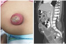
Instrumental diagnostics of cystic formation of the urachus is carried out using visualization methods: sonography (ultrasound) of the abdominal cavity and suprapubic region of the abdomen (bladder), computed tomography (CT), magnetic resonance imaging (MRI). Cystography is also performed.
The urachus cyst is visualized on ultrasound as an extra-abdominal mass with low echogenicity, located between the skin and the anterior abdominal wall, below the navel - along the midline of the abdomen. The contents of the inflamed cyst may appear heterogeneous.
Differential diagnosis
Differential diagnosis is carried out with a cyst of the mesentery or vitelline duct, a hernia of the umbilical or anterior abdominal wall, with a diverticulum of the bladder or ileum (Meckel's diverticulum), and with inflammation of the pelvic organs.
Who to contact?
Treatment urachus cysts
The presence of an asymptomatic urachal cyst does not usually require medical intervention. It is another matter if it increases in size or is accompanied by some symptoms. And the third situation is when the cyst becomes inflamed. And in the last two cases, treatment is necessary. [ 10 ]
And this is a surgical treatment, which consists of drainage and removal of the cyst (in case of small sizes – laparoscopically). [ 11 ], [ 12 ]
Prevention
To date, it is impossible to prevent congenital anomalies of the fetal urinary duct.
Forecast
The long-term prognosis for a urachus cyst, unless it becomes infected, is considered good.