Medical expert of the article

New publications
Spleen cyst in adult and child
Last reviewed: 12.07.2025

All iLive content is medically reviewed or fact checked to ensure as much factual accuracy as possible.
We have strict sourcing guidelines and only link to reputable media sites, academic research institutions and, whenever possible, medically peer reviewed studies. Note that the numbers in parentheses ([1], [2], etc.) are clickable links to these studies.
If you feel that any of our content is inaccurate, out-of-date, or otherwise questionable, please select it and press Ctrl + Enter.
Tumor-like formations in the form of cavities separated from the surrounding tissues can form in various organs, including the spleen. A splenic cyst (code D73.4 according to ICD-10) is considered rare, since it does not always manifest itself, and it is not so easy to detect it in the spleen covered with peritoneum. Often, a cyst of this localization is detected during an examination of the abdominal cavity for a completely different reason. [ 1 ]
Epidemiology
The widespread use of ultrasound and CT of abdominal organs has led to the fact that spleen cysts have become more frequently detected, and now they make up 1% of all diagnosed pathologies of this organ, and only 0.07% of diseases in the general population (according to some other data, 0.5-2%).
Statistically, non-parasitic cysts account for less than one-third of all splenic cysts, and most of these (almost two-thirds) are pseudocysts secondary to trauma. Only 10% of all non-parasitic splenic cysts are primary (congenital) cysts, which are most common in children and adolescents and rarely present clinically.[ 2 ]
Causes splenic cysts
Different types of splenic cysts have different causes of formation and histological features.
Non-parasitic cysts and parasitic cysts of the spleen (echinococcal) are distinguished. Non-parasitic cysts of the spleen can be epithelial (true) cysts or pseudocysts (false cysts). [ 3 ], [ 4 ]
Primary epithelial (epidermoid) cysts of the spleen are congenital, most often single (solitary) and quite large (with serous fluid inside). Their formation is associated with disorders of embryonic (intrauterine) development or genetically determined defects. Such a cyst of the spleen in a child or adolescent is the most common. [ 5 ], [ 6 ]
Most pseudocysts – the walls of which are made of fibrous tissue but are not lined with epithelium – arise from blunt abdominal trauma to the spleen with accumulation of blood (hematoma). Such a cyst in the spleen of an adult is usually filled with blood and dead cells. In a third of cases, its lining undergoes calcification, and then a calcified or calcified splenic cyst is determined. [ 7 ], [ 8 ]
A pseudocyst can be the result of infections, splenic infarction (for example, with thrombosis of the splenic artery), and with pancreatitis, such a cystic formation appears not only in the pancreas, but also in the spleen.
In addition to splenic infarction, a vascular splenic cyst may be caused by peliosis, the presence of small blood-filled cysts on the surface of the spleen.
A parasitic or echinococcal cyst of the spleen is formed as a result of infection with eggs and primary larvae of the parasitic tapeworm Echinococcus granulosus - echinococcus, which enter the body through the gastrointestinal tract and with the bloodstream - into the internal organs. The walls of these cysts are also often calcified. [ 9 ], [ 10 ]
Risk factors
The tendency to form cysts in the spleen in infants is observed in pathologies of pregnancy and prematurity of newborns; in adults - with increased destruction of blood platelets (thrombocytopenia), chronic viral infections, as well as with systemic lupus erythematosus, aplastic anemia, rheumatoid arthritis and other autoimmune diseases.
Risk factors for the development of splenic infarction, which can cause the formation of a vascular cyst, are associated with thrombi in the artery supplying the spleen with blood, atherosclerosis, systemic connective tissue diseases, and leukemia. The risk of developing peliosis increases with chronic alcoholism, HIV, tuberculosis, and the use of anabolic steroids and oral contraceptives. [ 11 ]
Pathogenesis
Any of the above causes can negatively affect the spleen and cause tissue damage.
When considering the pathogenesis of cyst formation in the spleen, experts emphasize its importance as one of the organs of the body's immune system, as well as its multifunctionality, including the deposition of erythrocytes and platelets, the production of leukocytes and antibodies, the metabolism of hemoglobin from spent erythrocytes, phagocytosis and filtration of blood (including from apoptosis products and pathological necrosis and toxic substances).
Researchers have not yet definitively determined how primary (congenital) cysts form in the spleen, but have suggested several versions. [ 12 ]
The formation of the spleen in the dorsal part of the mesentery from the mesodermal mesenchyme (with the participation of hematopoietic stem and dendritic cells) begins at the beginning of the second month of pregnancy, and until its completion the spleen is an organ of hematopoiesis, synthesizing erythrocytes.
The characteristic structure of the organ (lobules, trabeculae, parenchyma, venous system) is formed from the 15th week of gestation, and from approximately the 18th-19th week the stage of accumulation and differentiation of lymphocytes (T-cells) begins. [ 13 ]
So, the formation of cysts can be the result of the introduction of cells of the mesothelial membrane of the peritoneum into the splenic grooves of the fetus (and their metaplasia) or the inclusion of the endoderm of the internal germ layer into the lymphatic space or pulp of the forming organ.
The mechanism of development of an echinococcal cyst is caused by parasitic invasion: entering the spleen tissues with the bloodstream, the primary larvae of the tapeworm Echinococcus granulosus are transformed into the next stage - a finna, which is a capsule covered with a shell for further development of the parasite. Around these capsules, a parasitic cyst of the spleen or liver is formed. [ 14 ]
Symptoms splenic cysts
When a small splenic cyst is accidentally detected, most patients have no symptoms. However, when it is larger, the first signs may be discomfort on the left side of the hypochondrium and a painless mass in the upper left abdomen (detectable by palpation in a third of patients).
In addition, the following may appear: belching, rapid satiety when eating, aching pain in the left side, nausea and sometimes vomiting after eating, flatulence, diarrhea.
Also, during examination, swelling of the spleen and splenomegaly may be noted, especially if it is a parasitic cyst. Also, with an echinococcal cyst, there is general weakness and a slight increase in temperature.
A congenital cyst of the spleen in the fetus can be detected during a prenatal ultrasound, starting from the 20th week of pregnancy. A larger congenital cyst in the spleen of a newborn can be felt during a palpatory examination and, if enlarged, cause symptoms such as vomiting and intestinal upset. Most often, this is a single or solitary cyst of the spleen in newborns.
Read also:
Complications and consequences
What is dangerous about a spleen cyst? Usually, it does not cause complications, but the main negative consequences of this formation include:
- bleeding into the cyst "sac", which can lead to damage to the integrity of its walls;
- rupture of a splenic cyst with hemorrhage and spread of its contents into the abdominal cavity (for cysts larger than 5 cm, the risk is 25%), which may result in symptoms of acute abdomen and the development of peritonitis;
- infection of the cyst with suppuration, leading to intoxication of the body;
- spread of parasites from the echinococcal cyst to other organs.
Experts do not exclude the possibility of (extremely rare) malignant transformation of the cells of the secondary cyst membrane.
Diagnostics splenic cysts
Typically, diagnosis of a splenic cyst begins with the patient's history and requires a thorough clinical examination.
Blood tests: general clinical and biochemical, for antibodies (IgG) to echinococcus, for serum tumor markers (CEA, CA 19-9).
The main role is played by instrumental diagnostics: ultrasound, CT and/or MRI.
Congenital splenic cysts on ultrasound have the appearance of an anaechogenic mass with smooth walls. Epidermoid cysts have a complex structure with irregularities and thickness of the posterior walls due to epithelial peripheral trabeculae and internal echo from blood clots. See more - Ultrasound signs of spleen pathology
Currently, splenic cysts are known as a rare clinical condition with an incidence of 0.07% in the general population. Based on the presence or absence of cellular epithelial lining, these cysts are divided into primary (true) and secondary (false) cysts. Primary cysts are divided into parasitic (60%) and non-parasitic cysts depending on their etiology. Non-parasitic cysts are usually congenital. These cysts present mainly in young adults and are located in the upper pole of the spleen. [ 15 ]
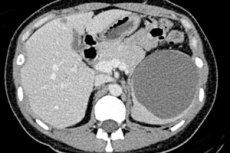
A splenic cyst is visualized in more detail on CT, therefore, performing a computed tomography of the spleen helps to determine many parameters of the cystic formation and make a more accurate diagnosis. [ 16 ]
Thus, by localization, there may be a cyst of the upper pole of the spleen (extremitas anterior), protruding forward above the colon; a cyst of the posterior pole (extremitas posterior) or on the inner part - in the area of the hilum of the spleen (hilum lienis). And with a deeper location - in its pulp or pulp (pulpa splenica) - a cyst in the parenchyma of the spleen is diagnosed.
The spleen is an encapsulated organ, and a subcapsular splenic cyst forms beneath the fibrous membrane (tunica fibrosa) of the organ.
In addition, a multilocular or multi-chamber cyst of the spleen often forms, and most often this is an echinococcal cyst.
Differential diagnosis
Differential diagnosis of cysts in the spleen includes its abscess, hemangioma, splenoma, lymphangioma, lymphoma, plasmacytoma, reciculo- and liposarcoma, teratoma. [ 17 ]
Treatment splenic cysts
It should be borne in mind that there is no medicine that can “dissolve” a cystic formation. Therefore, the treatment of a cyst with a diameter of more than 4 cm is surgical. [ 18 ]
Depending on the clinical situation, surgical treatment is carried out using methods such as:
- percutaneous aspiration of contents - laparoscopic puncture of splenic cyst; [ 19 ], [ 20 ]
- sclerotherapy of the cyst cavity with ethyl alcohol (after puncture removal of its contents);
- marsupialization (incomplete removal of the cyst mucosa, cystostomy);
- resection, that is, removal of the cyst;
- removal of the affected part of the spleen while preserving at least 30% of its parenchyma. [ 21 ]
However, in the case of multiple cysts, large cysts in the splenic hilum or in the parenchyma, or cysts with dense vascular adhesions to surrounding tissues, specialists consider open or laparoscopic splenectomy to be the method of choice. [ 22 ]
If the cyst is no more than 3 cm, its condition is monitored with annual ultrasound imaging.
Prevention
There is no way to prevent most spleen cysts from forming.
Forecast
The prognosis for the vast majority of cysts is good, but a splenic cyst larger than 5 cm in diameter has a high risk of rupture, which can lead to life-threatening intra-abdominal bleeding.