Medical expert of the article

New publications
Placenta maturation: late, premature
Last reviewed: 04.07.2025

All iLive content is medically reviewed or fact checked to ensure as much factual accuracy as possible.
We have strict sourcing guidelines and only link to reputable media sites, academic research institutions and, whenever possible, medically peer reviewed studies. Note that the numbers in parentheses ([1], [2], etc.) are clickable links to these studies.
If you feel that any of our content is inaccurate, out-of-date, or otherwise questionable, please select it and press Ctrl + Enter.
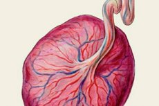
Maturation of the placenta is one of the important stages of the development of a healthy child in the future, since the placenta ensures the life of your baby. It is necessary to understand how the placenta matures under normal conditions, and what its functions are, in order to know what pathological changes and their signs may be.
What is the placenta for?
The function of the human placenta is of interest from both a scientific and clinical point of view. Studying the human placenta is very difficult because the methodology carries unacceptable risks for both mother and fetus.
The placenta plays a vital role in all trimesters of pregnancy and ensures that your baby develops safely. It performs many functions, such as:
- The placenta primarily functions to provide adequate nutrition to your baby. Before blood from you gets to your baby, it travels through the placenta to reach the umbilical cord that connects you to your baby. The placenta is the only organ in the body with two separate blood supplies, each coming from a different body. Because the placenta is a temporary organ, the blood flows change quickly with each stage of pregnancy.
The uteroplacental circulation is a low-resistance system for the maternal organism. Uterine arterial blood flow in the nonpregnant state averages 1% to 2% of the maternal cardiac output. During pregnancy, uterine blood flow increases rapidly until it reaches 17% of the maternal cardiac output.
The fetal placental circulation receives approximately two-thirds of the total fetal cardiac output. This high flow rate is important in transporting oxygen and nutrients from the mother to the fetus and is supported by a number of anatomical differences in the fetal circulation. Because the fetal lung has no respiratory function, high vascular resistance is maintained in this organ by the mechanical effects of the undilated alveoli on the vessel walls and the vasoconstrictor effect of the low oxygen tension that prevails in fetal blood. These two factors combine to shunt approximately two-thirds of the right ventricular output from the lungs to the systemic circulation via the ductus arteriosus.
This is one of the most important functions of the placenta.
- Another important function that the placenta performs is that it acts like a kidney; it filters the blood to remove harmful substances that may be dangerous to your baby's health.
- The placenta also serves as the baby's lungs and allows oxygen to pass to your baby.
- The placenta returns your baby's biological waste into mom's circulatory system, which is later removed from your body through urine.
- Throughout pregnancy, the placenta maintains the fundamental role of all biological membranes (i.e. selective permeability). With particles such as blood cells and macromolecules, the transfer is greatly restricted, providing a “placental barrier.” At the other end of the spectrum, the transfer of many essential nutrients is accelerated by various transport mechanisms. This will protect your baby from potential infections by separating the blood from your baby, acting as a filter.
- Many hormones are produced from the placenta in your body with maximum amounts of lactose, which ensures sufficient glucose levels in your blood to allow it to be distributed to the baby.
- The placenta also breaks down food particles you eat to ensure your baby is properly nourished.
- It takes the oxygen you breathe in to disperse it into your blood to help it reach your baby's circulatory system by passing it through the umbilical cord. This is one of the important functions that the placenta performs, as it prevents the possibility of your baby inhaling amniotic fluid, which can be disastrous.
- The placenta secretes a huge amount of female hormones, such as progesterone and estrogen, which provide tone to the uterus, growth of the placenta, delay of the next ovulation and support of the pregnancy itself. It also paves the way for the preparation of maternal tissues and the uterus for the birth of the baby.
- During the stages of pregnancy, the placenta moves as the uterus expands and grows. It is the general function of the placenta to remain in the early stages of pregnancy, but in the later stages of pregnancy, it moves to the top of the uterus to open the cervix for the birth of the baby.
Normal maturation of the placenta
The placenta is the fastest growing organ in the human body. The placenta grows from one cell to approximately 5 x 10 to the 10th power cells in 38 weeks. Implantation of the fertilized egg occurs seven to ten days after conception. The layer of cells that form the surface of the embryo develops into the chorionic membrane, and the cytotrophoblast cells are derived from it. Trophoblast cells are multinucleated aggregates of cytotrophoblast cells and are continually being formed from them. These cells, plus the villi, are the characteristic and unique features of the future human placenta.
Venous sinuses within the endometrium penetrate the trophoblast cells very early. Within a few days, lacunae develop, surrounded by syncytial cells and filled with maternal venous blood and tissue fluid. The maternal spiral arterioles are destroyed on the 14th or 15th day, and maternal arterial blood enters the developing space. Fetal vessels form in situ within the mesenchymal cores, and the resulting villi are called tertiary villi. Around the 17th day after conception, both fetal and maternal blood vessels are functional, and true placental circulation is established. This underlies the formation of the future placenta.
Fetal and maternal vascularization of the placenta is complete by days 17–20, and fetal embryonic red blood cells can be detected within fetal vessels after day 21 postconception. The placenta continues to grow in thickness and girth until the end of the fourth month. The increase in placental thickness is due to an increase in the length and size of the villi with an accompanying widening of the intervertebral space. There is no noticeable increase in thickness after the fourth month, but growth in girth continues throughout most of pregnancy.
The human placenta is a hemochronic placenta, which means that the maternal blood is in direct contact with the embryonic trophoblast. The maternal blood circulates freely in space. The functional unit of the placenta can be considered a villus, it is here that the exchange of substances between the mother and the fetus occurs at the molecular level. Therefore, the development of placental villi is the basis for the correct development and maturation of the placenta.
In early placentation, each placental villus goes through a similar initial developmental program. In late placentation, the villi differentiate morphologically into a limited range of functional villus changes that reflect their specialization. The main initial contribution consists of the trophoblast membrane that surrounds the embryo and then, through the development of extraembryonic mesoderm and differentiation of blood vessels, performs its function.
There are three main types of trophoblast cells: villous cytotrophoblasts, extravillous cytotrophoblasts, and syncytiotrophoblasts, which are formed by the fusion of villous cytotrophoblasts.
The syncytiotrophoblast layer forms the epithelial covering of the entire villous tree. These cells are multinucleated, terminally differentiated syncytium formed by the fusion of cytotrophoblast precursor cells. Differentiation is regulated by human chorionic gonadotropin, and fusion of cytotrophoblast cells continues during placental development.
Cellular parts derived from syncytiotrophoblasts (apoptotic nuclei and microparticles) can be shed into the maternal blood.
Mesenchymal villi are continuously formed from trophoblastic villi throughout pregnancy and are considered to be the basis for the growth and differentiation of villous trees. They will form the basis of the functional unit of the future placenta.
Initially, primary villi are formed. Thus, in the second week of placental development, the first stage of development of chorionic villi, trophoblastic membrane cells (syncytotrophoblasts and cytotrophoblasts), which form finger-like extensions into the maternal decidua, occurs.
Secondary villi develop in the third week – this is the second stage of development of the chorionic villus. At this time, the extraembryonic mesoderm turns into villi and covers the entire surface of the chorionic sac.
Tertiary villi are formed in the 4th week - this is already the third stage of development of the chorionic villus. At this stage, the mesenchyme differentiates into blood vessels and cells, forming an arteriocapillary network.
In the first two trimesters, tertiary villi are precursors of immature intermediate villi, whereas in the last trimester, mesenchymal villi develop into mature intermediate villi. Immature intermediate villi formed during the first two trimesters are developmental steps in relation to stem villi.
Mature intermediate villi develop during the last trimester, producing numerous terminal villi. Terminal villi are not active outgrowths caused by trophoblast proliferation, but rather passive protrusions caused by capillary twisting due to excessive longitudinal growth of fetal capillaries in mature intermediate villi.
The development of the placenta corresponds to the gestational age. At 4-5 weeks, a complex network of cords and vessels with redundant connections is initially formed. This network contains mainly cords already connected together. The vessels and cords are connected to each other without interruptions.
At 6-7 weeks, the villi, in which the capillary network of vessels and cords predominates, form the basis of the villus.
At 8-9 weeks, the villi have two large centralized vessels that are surrounded and connected to the peripheral capillary network. The capillary network contains vessels with a lumen in close contact with the overlapping trophoblastic layer. This ensures the further development of the vascular network of the placenta.
The maturation period of the placenta begins from the very first moment of development of the first villi and lasts no less than thirty weeks.
The norm of placental maturation has successive stages at the macroscopic level. Knowledge and distinction of such stages is very important for assessing the condition of the fetus and the functional development of the placenta itself. The following degrees of placental maturation are distinguished by weeks:
0 (zero) degree is characterized by the formation of a clear, correct structure, in which all lobes of the placenta are fully formed. In this case, each villus has reached the final stage of growth, it has the weight of a cell and vessels necessary for gas exchange. This degree is characteristic of the complete completion of the formation of the placenta, and it should normally be at the thirtieth week of pregnancy. Such a placenta at this stage can provide all the functions and needs of the baby at this stage of gestation.
- The degree is characterized by a change in the homogeneity of the placenta tissue and the formation of areas of different echogenicity. This is a normal process and it indicates the sequential development of different areas of the placenta. This degree is typical for the thirtieth to thirty-third week of pregnancy. There may be a variation of one week.
- The stage develops at the thirty-fourth to thirty-seventh week. At the same time, the chorionic plate becomes tortuous, echogenic areas appear in greater quantities. This stage is considered the most mature and functionally active. At the same time, the thickness of the placenta at this stage is from 29 to 49 millimeters. Such functional activity of the placenta allows the child to receive maximum nutrients to store them for the period of labor.
- the degree of maturity indicates the complete readiness of the placenta for the process of physiological birth. At the same time, the processes of division of the placenta and the formation of its apical and distal ends begin. This degree develops just before the birth and should be observed at least at 39 weeks.
Causes placenta maturation disorders
Although the placenta is a reliable organ, various factors can affect the health of the placenta during pregnancy stages, making the pregnant mother prone to pregnancy and fetal pathology. Although some of these problems can be addressed and modified, the main causes of placental maturation disorders are as follows:
- Trauma to the abdomen of a pregnant woman due to a fall or any other type of impact.
- Blood clotting problems: Certain medical conditions can interfere with the blood's ability to clot, which can disrupt uterine and placental blood flow.
- High blood pressure levels can significantly damage the health of the placenta. Because the change in pressure changes the flow and strength of blood in the placenta, so the villi of the placenta may not develop as they should.
- High maternal age: this is a risk factor for disruption of placental formation, since at this age the processes of cell differentiation and division are reduced.
- Multiple pregnancies: Mothers who are pregnant with twins or triplets often develop a weak placenta because all the nutrients need to be distributed properly.
- Premature rupture of membranes: The fluid-filled membrane known as the amniotic sac can rupture early, disrupting the structure and leading to complications.
- A pregnant woman who has a history of placental problems in a previous pregnancy has risk factors for developing the same problem with each subsequent pregnancy.
- Uterine surgery: Any previous surgery on the uterus may result in implantation failure and subsequent disruption of placenta formation.
 [
[ Pathogenesis
The pathogenesis of abnormal maturation can be observed in several different settings. Accelerated premature maturation of the placenta, i.e. premature formation of terminal villi, can be considered as a reaction or adaptation of the placenta to reduced maternal-placental perfusion. Histologically, this can be recognized by a decrease in villus diameter and accelerated formation of syncytiovascular membranes.
Late maturation of the placenta, when labor begins and the placenta is not yet mature, indicates that factors acted intrauterine that stopped the maturation of the placenta. Therefore, at a late gestational age, the placenta is not yet mature enough to provide normal blood flow. Such late maturation can be observed in several different clinical situations. It occurs in women with diabetes on the maternal side. It can be observed in connection with congenital or chromosomal abnormalities, with chronic vilitis (inflammation of the villi) of unknown etiology.
Symptoms placenta maturation disorders
A woman cannot feel the symptoms of premature maturation of the placenta. Since the placenta is an organ that ensures the life of the fetus, the first signs of impaired placental maturation will appear precisely from the side of changes in the functional activity of the fetus. A woman may notice that the fetus has begun to move less or, and such changes will not be reflected in her well-being.
The diagnosis of accelerated placental maturation is mainly based on additional research methods. Instrumental diagnostics of placental maturation allows determining its thickness and the nature of the structure of the chorionic plate. If, for example, the third degree of placental maturity is determined at the thirtieth week of pregnancy, this can be considered accelerated or premature maturation. This pathology is confirmed by ultrasound data. Since at this stage the child is not yet ready for birth, such aging of the placenta can have consequences. Complications can develop, since with such accelerated maturation of the placenta, calcifications are formed in the placenta and blood circulation processes are disrupted. What is the threat of premature maturation of the placenta? This can lead to premature birth, or, if the process is formed gradually, chronic fetal hypoxia develops.
Clinical signs of late maturation of the placenta are also difficult to diagnose without tests and instrumental methods. If before birth at the thirty-seventh and thirty-ninth week the degree of maturity of the placenta is less than the second, then this indicates a delay or late maturation. In such cases, the development of uterine and placental vascular anastomoses is delayed, as well as insufficient hormonal function of the placenta. The consequences and complications of this condition can be serious, including congenital anomalies in the child.
Diagnosis of placental maturation anomalies is primarily an ultrasound examination. Ultrasound signs of premature placental maturation are a thickening of the placenta width of more than 35 millimeters, the appearance of calcifications or inclusions in the thickness of the placenta, as well as increased waviness of the chorionic plate.
Tests that can confirm a particular diagnosis of pathology are not specific. In certain cases, additional tests are required to identify the cause that led to such disorders.
Differential diagnosis
Differential diagnostics of placental maturation disorders should be performed at the stage of ultrasound diagnostics. It should exclude infectious lesions of the placenta, which require immediate treatment at the stage of detection.
Treatment placenta maturation disorders
What to do with premature maturation of the placenta? Treatment of placental maturation pathologies has many conditions. Here, an important question is how pronounced the changes are. If premature maturation is observed only at one degree, then drug correction and expectant therapy are possible against the background of monitoring the condition.
Drug therapy is used to improve uterine circulation, which suffers due to these pathologies.
Curantil is used as a means of pathogenetic therapy in such cases. The drug improves the rheological properties of blood and prevents the formation of blood clots. The method of administration is oral. Dosage - starting from 75 milligrams per day, if necessary, the dose can be increased. Side effects can be in the form of postpartum bleeding if the drug is used for a long time.
Other medications used in the treatment of pregnant women with premature placenta have not been proven effective, so they should be used with caution.
If we talk about folk methods of treatment, as well as homeopathic remedies, they can be used only on the doctor's recommendations. Considering the possible risks for the fetus, when there are already violations of placental maturation, it is important to maintain maximum functionality until the very period of labor. Therefore, folk remedies should be used carefully.
Prevention
Prevention of placental maturation disorders consists of general measures that ensure a healthy pregnancy. It is important to plan pregnancy and treat diseases before it occurs. If a woman has chronic pathologies, they can cause placental formation disorders, so such pathologies need to be treated in a timely manner.
Forecast
The prognosis for the birth of a child with delayed maturation of the placenta or with accelerated maturation of the placenta is generally favorable. Due to the disruption of uterine blood circulation, there may be risks of giving birth to children with intrauterine growth retardation. More serious congenital anomalies with these pathologies are very rare.
Placental maturation is a long and very important process for the development of the baby. This organ has a unique structure that provides many basic functions for the fetus. Therefore, any violations of placental maturation should be prevented, because they can have consequences.