Medical expert of the article
New publications
The ulnar nerve
Last reviewed: 07.07.2025

All iLive content is medically reviewed or fact checked to ensure as much factual accuracy as possible.
We have strict sourcing guidelines and only link to reputable media sites, academic research institutions and, whenever possible, medically peer reviewed studies. Note that the numbers in parentheses ([1], [2], etc.) are clickable links to these studies.
If you feel that any of our content is inaccurate, out-of-date, or otherwise questionable, please select it and press Ctrl + Enter.
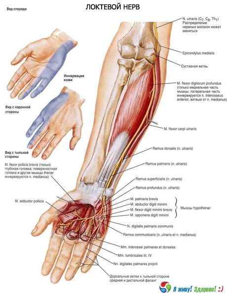
The ulnar nerve (n. ulnaris) departs from the medial cord of the brachial plexus. It consists of fibers of the anterior branches of the eighth cervical - first thoracic (CVIII-ThI) spinal nerves. Initially, the ulnar nerve is located next to the median nerve and slightly medial to the brachial artery. In the middle third of the arm, the nerve deviates to the medial side, then pierces the medial intermuscular septum of the arm and goes down to the posterior surface of the medial epicondyle of the humerus. On the arm, the ulnar nerve does not give off branches. Then the ulnar nerve gradually shifts to the anterior surface of the forearm, where it initially passes between the muscle bundles of the initial part of the flexor carpi ulnaris. Below, the nerve is located between the flexor carpi ulnaris medially and the flexor digitorum superficialis laterally. At the level of the lower third of the forearm, it runs in the ulnar groove of the forearm, next to and medial to the arteries and veins of the same name. Closer to the head of the ulna, the dorsal branch (r. dorsalis) of the ulnar nerve departs from it, which on the back of the hand runs between this bone and the tendon of the ulnar flexor of the wrist. On the forearm, the muscular branches of the nerve innervate the ulnar flexor of the wrist and the medial part of the deep flexor of the fingers.
The dorsal branch of the ulnar nerve on the back of the hand divides into five dorsal digital branches. These nerves innervate the skin of the back of the hand on the ulnar side, the skin of the proximal phalanges of the fourth, fifth and ulnar side of the third finger.

The palmar branch (r. palmaris) of the ulnar nerve, together with the ulnar artery, passes to the palm through a gap in the medial part of the flexor retinaculum, on the lateral side of the pisiform bone. Near the uncinate process of the uncinate bone, the palmar branch divides into a superficial and a deep branch. The superficial branch (r. superficialis) is located under the palmar aponeurosis. From it, a branch initially goes to the short palmaris muscle. Then it divides into the common palmar digital nerve (n. digitalis palmaris communis) and the proper palmar nerve. The common palmar digital nerve passes under the palmar aponeurosis and in the middle of the palm divides into two proper palmar digital nerves. They innervate the skin of the facing sides of the fourth and fifth fingers, as well as the skin of their dorsal surfaces in the region of the middle and distal phalanx. The palmar digital nerve (n. digitalis palmaris proprius) innervates the skin of the ulnar side of the little finger.
The deep branch (r. profundus) of the ulnar nerve initially accompanies the deep branch of the ulnar artery. This branch passes between the muscle that abducts the little finger medially and the short flexor of the little finger laterally. Then the deep branch deviates to the side, runs obliquely between the bundles of the muscle that abducts the little finger, under the distal parts of the flexor tendons of the fingers, located on the interosseous palmar muscles. The deep branch of the ulnar nerve innervates all the muscles of the eminence of the little finger (the short flexor of the little finger, the abductor and opposer muscles of the little finger), the dorsal and palmar interosseous muscles, as well as the adductor muscle of the thumb and the deep head of the short flexor of the thumb, the 3rd and 4th lumbrical muscles, bones, joints and ligaments of the hand. The deep palmar branch is connected by communicating branches with the branches of the median nerve.
How to examine?

