Medical expert of the article

New publications
Brachial plexus
Last reviewed: 04.07.2025

All iLive content is medically reviewed or fact checked to ensure as much factual accuracy as possible.
We have strict sourcing guidelines and only link to reputable media sites, academic research institutions and, whenever possible, medically peer reviewed studies. Note that the numbers in parentheses ([1], [2], etc.) are clickable links to these studies.
If you feel that any of our content is inaccurate, out-of-date, or otherwise questionable, please select it and press Ctrl + Enter.
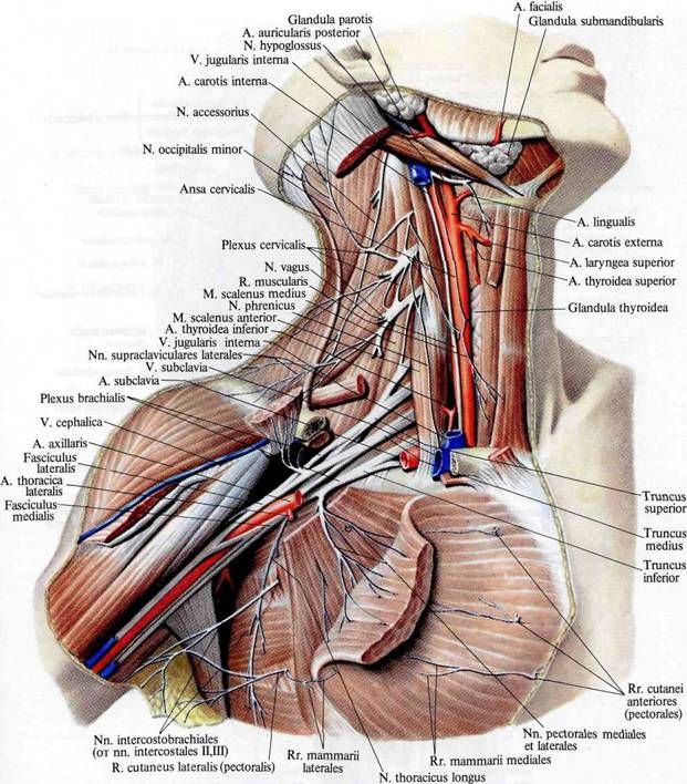
The brachial plexus (plexus brachialis) is formed by the anterior branches of the four lower cervical (CV-CVIII) spinal nerves. Based on topographic features, the plexus is divided into supraclavicular and infraclavicular parts (pars supraclavicularis et pars infraclavicularis). Initially, the brachial plexus is located in the interscalene space (supraclavicular part), where the upper, middle and lower trunks of the brachial plexus are distinguished. From the interscalene space, these trunks exit into the large supraclavicular fossa (scapulotrapezoid triangle). At the level of the clavicle and below, the trunks of the brachial plexus form three bundles (subclavian part) surrounding the axillary artery in the axillary cavity. In relation to the axillary artery, these are the medial, lateral and posterior bundles (fasciculi medialis, lateralis, posterior) of the brachial plexus. The brachial plexus receives connecting branches from the middle cervical ganglion of the sympathetic trunk of its side. Short and long branches depart from the brachial plexus. Short branches come mainly from the supraclavicular part of the brachial plexus. They innervate the bones and soft tissues of the shoulder girdle. Long branches of the brachial plexus depart from the subclavian part of the brachial plexus and innervate the free part of the upper limb.
Short branches of the brachial plexus
The short branches of the brachial plexus include the dorsal (posterior) nerve of the scapula, the long thoracic, subclavian, suprascapular, subscapular, thoracospinal, lateral and medial thoracic nerves, and the axillary nerve. Muscular branches are also considered short branches of the brachial plexus; they innervate the scalene muscles and the splenius muscle of the neck.
- The dorsal nerve of the scapula (n. dorsalis scapulae) originates from the anterior branches of the fourth and fifth cervical spinal nerves. The nerve passes along the anterior surface of the muscle that lifts the scapula, then between the middle and posterior scalene muscles and branches into the large and small rhomboid muscles and the muscle that lifts the scapula.
- The long thoracic nerve (n. thoracicus longus) originates from the anterior branches of the fifth and sixth spinal nerves (CV-CVI), goes behind the brachial plexus. Then the nerve is located between the subscapularis and anterior serratus muscles, goes down between the lateral thoracic artery in front and the thoracospinal artery behind. It innervates the anterior serratus muscle.
- The subclavian nerve (n. subclavius) is formed by the anterior branch of the fifth spinal nerve. The nerve takes the shortest route down the outer edge of the anterior scalene muscle to the subclavian muscle. Often, the subclavian nerve gives off a branch to the phrenic nerve.
- The suprascapular nerve (n. suprascapularis) is formed by the anterior branches of the fifth and sixth spinal nerves. It is separated directly from the superior bundle of the brachial plexus. At first, the nerve passes near the superior edge of the brachial plexus under the trapezius muscle and the inferior belly of the omohyoid muscle. Then, behind the clavicle, the nerve forms a bend laterally and posteriorly, passes into the supraspinous fossa through the notch of the scapula under its superior transverse ligament. Then, together with the transverse artery of the scapula, the suprascapular nerve passes under the base of the acromion into the infraspinous fossa. It innervates the supraspinatus and infraspinatus muscles and the capsule of the shoulder joint.
- The subscapular nerve (n. subscapularis) departs from the anterior branches of the fifth to seventh spinal nerves with two or three trunks and runs along the anterior surface of the subscapularis muscle. It innervates the subscapularis and teres major muscles.
- The thoracodorsal nerve (n. thoracodorsalis) is formed from the anterior branches of the fifth through seventh spinal nerves and runs downward along the outer edge of the scapula to the latissimus dorsi, which it innervates.
- The lateral and medial pectoral nerves (nn. pectorales lateralis et medialis) originate from the lateral and medial bundles of the brachial plexus (CV-ThI). The nerves go forward, pierce the clavipectoral fascia and end in the pectoralis major (medial nerve) and pectoralis minor (lateral nerve) muscles.
- The axillary nerve (n. axillaris) originates from the posterior cord of the brachial plexus (CV-CVIII). The nerve runs laterally and downwards along the anterior surface of the subscapularis muscle, then turns back. Together with the posterior circumflex humeral artery, the nerve passes through the quadrilateral opening and exits onto the dorsal surface of the shoulder. Then the nerve enters the deltoid muscle from the lateral surface of the surgical neck of the humerus, giving off a small branch to the teres minor muscle and the capsule of the shoulder joint. The terminal branch of the axillary nerve is the superior lateral cutaneous nerve of the arm (n. cutaneus brachii lateralis superior), which exits under the skin between the posterior edge of the deltoid muscle and the long head of the triceps brachii and innervates the skin above the deltoid muscle and in the lateral part of the shoulder.
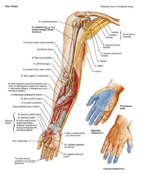
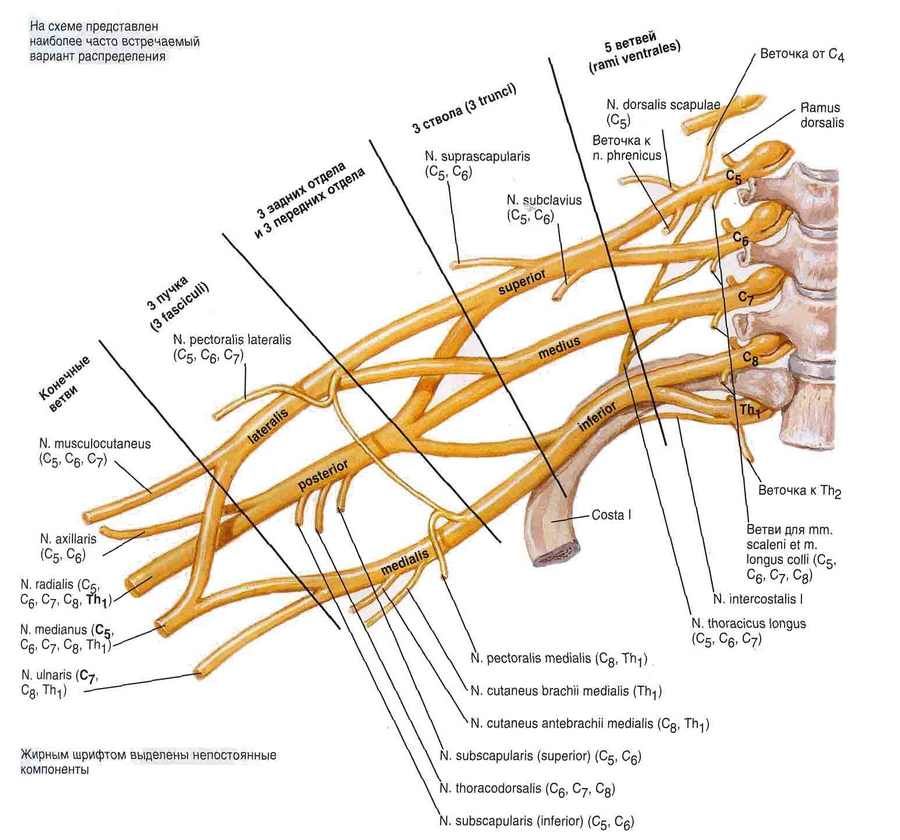
Long branches of the brachial plexus
The long branches of the brachial plexus originate from the lateral, medial, and posterior bundles of the subclavian part of the brachial plexus. Among the long branches, the following are distinguished: the musculocutaneous, median, ulnar nerves, the medial cutaneous nerve of the arm, the medial cutaneous nerve of the forearm, and the radial nerve.
- The musculocutaneous nerve (n. musculocutaneus) departs from the lateral cord of the brachial plexus. This nerve is formed by the anterior branches of the fifth to eighth (CV-CVIII) cervical spinal nerves. The musculocutaneous nerve goes downward and laterally, pierces the coracobrachialis muscle and gives off branches to it. Initially, the nerve is located lateral to the median nerve, then separates from it downwards. On the shoulder, the musculocutaneous nerve passes between the brachialis and biceps brachii muscles, giving off muscular branches to them (rr. musculares). At the level of the elbow joint, lateral to the terminal part of the biceps tendon, the musculocutaneous nerve pierces the fascia of the arm and continues into the lateral cutaneous nerve of the forearm (n. cutaneus anteabrachii lateralis), which descends downward under the skin along the lateral side of the forearm. The lateral cutaneous nerve of the forearm innervates the skin of this region up to the eminence of the thumb.
- The median nerve (n. medianus) originates from the junction of the lateral and medial bundles of the brachial plexus formed by the fibers of the anterior branches of the sixth to eighth cervical and first thoracic (CVI-ThI) spinal nerves. Both bundles join at an acute angle in front of the axillary artery. On the shoulder, the median nerve initially passes in the same fascial sheath with the brachial artery, being located lateral to it. The projection of the median nerve corresponds to the location of the medial groove of the shoulder.
- The ulnar nerve (n. ulnaris) departs from the medial cord of the brachial plexus. It consists of fibers of the anterior branches of the eighth cervical - first thoracic (CVIII-ThI) spinal nerves. Initially, the ulnar nerve is located next to the median nerve and slightly medial to the brachial artery. In the middle third of the arm, the nerve deviates to the medial side, then pierces the medial intermuscular septum of the arm and goes down to the posterior surface of the medial epicondyle of the humerus.
- The medial cutaneous nerve of the arm (n. cutaneus brachii medialis) is formed by the fibers of the anterior branches of the eighth cervical and first thoracic spinal nerves (CVIII-ThI), departs from the medial cord of the brachial plexus and accompanies the brachial artery. At the base of the axillary cavity, the medial cutaneous nerve of the arm joins the lateral cutaneous branches of the second and third intercostal nerves and is called the intercostobrachial nerve (n. intercostobrachialis). Then the medial cutaneous nerve of the arm pierces the axillary and brachial fascia and branches in the skin of the medial side of the arm to the medial epicondyle of the humerus and the olecranon process of the ulna.
- The medial cutaneous nerve of the forearm (n. cutaneus antebrachii medialis) consists of fibers of the anterior branches of the eighth cervical - first thoracic (CVII-ThI) spinal nerves. It emerges from the medial cord of the brachial plexus and is adjacent to the brachial artery. Initially, the nerve is located deep on the shoulder, then pierces the fascia of the arm at the point where the medial saphenous vein of the arm enters one of the brachial veins. The branches of the medial cutaneous nerve of the forearm innervate the skin of the medial side of the lower arm and the back of the non-medial side of the forearm.
- The radial nerve (n. radialis) is a continuation of the posterior cord of the brachial plexus. It consists of fibers of the anterior branches of the fifth cervical - first thoracic (CV-ThI) spinal nerves.