Medical expert of the article

New publications
Radial nerve
Last reviewed: 07.07.2025

All iLive content is medically reviewed or fact checked to ensure as much factual accuracy as possible.
We have strict sourcing guidelines and only link to reputable media sites, academic research institutions and, whenever possible, medically peer reviewed studies. Note that the numbers in parentheses ([1], [2], etc.) are clickable links to these studies.
If you feel that any of our content is inaccurate, out-of-date, or otherwise questionable, please select it and press Ctrl + Enter.
The radial nerve (n. radialis) is a continuation of the posterior cord of the brachial plexus. It consists of fibers of the anterior branches of the fifth cervical - first thoracic (CV-ThI) spinal nerves. In terms of thickness, the radial nerve is the largest branch of the brachial plexus. It begins at the level of the lower edge of the pectoralis minor muscle. Initially, the nerve goes behind the axillary artery, then between the lateral and medial heads of the triceps brachii muscle it passes into the brachiomuscular (spiral) canal. Before entering this canal, the posterior cutaneous nerve of the arm (n. cutaneus brachii posterior) branches off from the radial nerve, goes backward, pierces the long head of the triceps brachii muscle and the fascia of the arm near the tendon of the deltoid muscle. The nerve innervates the skin of the posterolateral surface of the arm.
In the brachiomuscular canal, the posterior cutaneous nerve of the forearm (n. cutaneus antebrachii posterior) branches off from the radial nerve. This nerve initially goes together with the radial nerve, then passes between the lateral and medial heads of the triceps brachii. The nerve exits onto the back of the forearm and innervates the skin of its back side to the level of the wrist joint. On the shoulder, the radial nerve innervates the triceps brachii and the antecubital muscle.
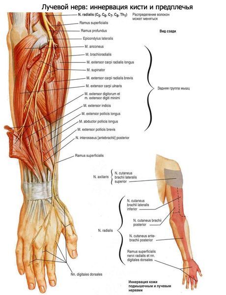
After leaving the brachial canal, the radial nerve pierces the lateral intermuscular septum of the shoulder and descends between the brachialis and the origin of the brachioradialis muscle. At the level of the elbow joint, the radial nerve divides into superficial and deep branches. The superficial branch (r. superficialis) of the radial nerve is thinner and longer than the deep branch of this nerve. At first, the superficial branch goes down under the brachioradialis muscle, then between the brachioradialis muscle and the long radial extensor of the wrist. In the lower third of the forearm, the superficial branch is located subcutaneously, gradually deviates in the lateral direction, then passes to the back of the forearm between the radius and the tendon of the brachioradialis muscle. At a distance of 4-5 cm above the level of the styloid process of the radius, this branch gives off branches to the skin of the dorsal and lateral sides of the base of the thumb and divides into five dorsal digital nerves (nn. digitales dorsales). Two of these nerves are directed to the radial and ulnar sides of the thumb, innervating its skin from the dorsal side. The remaining three digital dorsal nerves branch out in the skin of the second finger and the radial side of the third finger at the level of their proximal phalanges. The skin of the dorsum of the middle and distal phalanges of the second and third fingers is innervated by the palmar digital nerves of the median nerve. The deep branch (r. profundus) of the radial nerve pierces the supinator muscle, gives off muscle branches to it and to the short radial extensor of the wrist. Near the radius, the deep branch passes to the back of the forearm, where it gives off muscle branches to the remaining muscles of the back of the forearm. The longest of these branches is the posterior interosseous nerve (n. interosseus posterior). It passes between the superficial and deep layers of muscles on the back of the forearm, innervating the interosseous membrane and the muscles located nearby.
 [
[ How to examine?