Medical expert of the article

New publications
Sialography
Last reviewed: 06.07.2025

All iLive content is medically reviewed or fact checked to ensure as much factual accuracy as possible.
We have strict sourcing guidelines and only link to reputable media sites, academic research institutions and, whenever possible, medically peer reviewed studies. Note that the numbers in parentheses ([1], [2], etc.) are clickable links to these studies.
If you feel that any of our content is inaccurate, out-of-date, or otherwise questionable, please select it and press Ctrl + Enter.
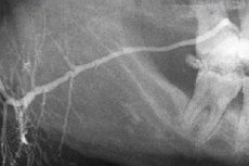
Methodology for performing sialography
Sialography involves examining the ducts of the major salivary glands by filling them with iodine-containing preparations. For this purpose, water-soluble contrast or emulsified oil preparations (dianosyl, ultra-liquid lipoiodinol, etiidol, mayodil, etc.) are used. Before administration, the preparations are heated to a temperature of 37-40 °C to prevent cold spasm of the vessels.
The study is conducted with the aim of diagnosing mainly inflammatory diseases of the salivary glands and salivary stone disease.
A special cannula, a thin polyethylene or non-latonic catheter with a diameter of 0.6-0.9 mm or a blunt and slightly bent injection needle are inserted into the opening of the excretory duct of the examined salivary gland. After bougienage of the duct, the catheter with a mandrel, inserted into it to a depth of 2-3 cm, is tightly grasped by the walls of the duct. For examination of the parotid gland, 2-2.5 ml is introduced, for the submandibular gland - 1-1.5 ml of contrast agent.
Radiography is performed in standard lateral and direct projections; sometimes axial and tangential images are taken.
When contrasting several salivary glands simultaneously, panoramic tomography (pantomosialography) is preferable, as it allows obtaining a sufficiently informative picture in one image with low radiation exposure to the patient.
Analysis of images taken 15-30 minutes later allows us to judge the function of the salivary glands. Citric acid is used to stimulate salivation.
Sialography in combination with CT is successfully used to differentiate benign and malignant tumors of the parotid salivary gland.
In recent years, ultrasound and functional digital subtraction sialography have been used to diagnose salivary gland diseases. Contrast agents are introduced into cystic formations by puncturing the cyst wall. After the contents are aspirated, a heated contrast agent is introduced into the cavity. Radiographs are taken in two mutually perpendicular projections.
Oil (iodolipol, lipiodol, etc.) or water-soluble (76% verografin solution, 60% urografin solution, omnipaque solution, trasograph, etc.) preparations are used as a contrast agent. Water-soluble preparations are advisable to use in cases where there is a risk of the substance getting beyond the salivary gland (in patients with Sjogren's syndrome, with duct strictures, malignant tumors) and in cases of contraindications to long-term retention of iodine preparations in the ducts (in patients who are to undergo radiation therapy). The contrast agent is slowly injected through the duct into the gland until the patient feels a feeling of distension in it, which corresponds to filling of the ducts of the first to third orders. To fill the ducts of the unchanged parotid gland, 1-2 ml of an oil or 3-4 ml of a water-soluble preparation is required. To fill the ducts of the submandibular gland - 1.0-1.5 ml and 2.0-3.0 ml, respectively.
Sialography of the salivary glands is performed only during the period of remission of the process. Otherwise, the course of sialadenitis may worsen.
The most complete picture of the structure of the parotid gland is obtained on a sialogram in the lateral projection. On a sialogram of the submandibular glands in the lateral projection, the submandibular duct is determined at the level of the body of the lower jaw, the gland with its upper pole is superimposed on the angle of the lower jaw, the greater part is determined below its base.
Pantomosialography
This is sialography with simultaneous contrasting of two parotid, two submandibular or all four salivary glands followed by panoramic tomography. This method is indicated in the same cases as sialography. Simultaneous examination of paired glands allows to detect clinically hidden inflammatory process in the paired gland.
The description of the sialogram is made according to the following scheme. In relation to the parenchyma of the gland, the following is established:
- how the image is revealed (good; unclear but uniform; unclear and uneven; not revealed);
- presence of a filling defect in the ducts;
- the presence of cavities of different diameters;
- clarity of cavity contours.
When examining the ducts, the following is determined:
- narrowing or widening of the IV order ducts (uniform, uneven);
- dilation of the parotid or submandibular ducts (uniform, uneven);
- mixing or interruption of ducts;
- clarity of duct contours (clear, fuzzy).
Digital sialography
This is sialography, which is performed on special devices (usually with digital information), allowing to obtain a more contrasting image and analyze it in the dynamics of filling of the gland and evacuation of the contrast agent.
Digital subtraction sialography increases the diagnostic capabilities of sialography due to subtraction (subtraction of the surrounding background of bone and tissue formations) and the ability to visualize the filling and evacuation of the contrast agent in the dynamics of the study. The examination is carried out on X-ray machines with a digital attachment or on angiographs; the examination time is 30-40 s. An analysis of the duct system picture, the filling time and evacuation of the water-soluble contrast agent is performed.
Sialadenolymphography
The method was proposed by V.V. Neustroev et al. (1984) and Yu.M. Kharitonov (1989) for diagnostics of salivary gland diseases based on the study of their lymphatic apparatus (intra- and extraorgan lymphatic system). Using a syringe and needle, 4 ml of water-soluble or 2 ml of fat-soluble contrast agent are injected percutaneously into the parotid gland. Serial sialadenolmphography is performed after 5 and 20 min, 2 and 24 h. The authors indicated that the X-ray semiotics of chronic sialadenitis is associated with an uneven depleted pattern of intraorgan lymphatic vessels with preservation of the organ contours and regional lymph outflow. In tumors, a filling defect is determined.
Computerized sialtomography
The image is obtained on computer tomographs. Scanning begins from the level of the hyoid bone with a Gantry tilt of 5° for the submandibular and 20° for the parotid glands. 15 sections are taken with a step (thickness) of 2-5 mm. The resulting cross-section is topographic-anatomical, similar to Pirogov's. The method is indicated for diagnosing salivary stone disease and various types of salivary gland tumors.
Radionuclide methods of examination (radiosialography, scanning and scintigraphy) are based on the selective ability of glandular tissue to absorb radioactive isotopes I-131 or Technetium-99m (pertechnetate). These methods are practically harmless, since patients are administered indicator doses of a radiopharmaceutical with a radiation power 20-30 times less than during a conventional X-ray examination. The methods allow an objective assessment of the functional state of the secreting parenchyma regardless of the quality and quantity of secretion, and to conduct differential diagnostics between a tumor and inflammation of the salivary gland.
Radiosialography of the parotid glands (radioisotope sialometry) was developed by L.A. Yudin. The study involves recording the curves of the intensity of radioactive radiation over the parotid glands and heart after intravenous administration of pertechnetate (Tc-99m) at a dose of 7.4-11.1 MBq and allows for an objective assessment of their function. A radiosialogram of unchanged parotid glands normally consists of three curves: in the first minute, there is a sharp increase in radioactivity over the salivary glands, then a small rapid decline (the first vascular section of the curve). Then, over the course of 20 minutes, the radioactivity gradually increases. This section is called the concentration section. The increase in radioactivity stops or is less intense (plateau). This level of radioactivity corresponds to the maximum accumulation of the radiopharmaceutical (MAR). Normally, the MAR time is 22 ±1 min for the right and 23+1 min for the left parotid gland. After 30 minutes, stimulation of salivation with sugar leads to a sharp (within 3-5 minutes) drop in radioactivity, and this section is called the excretory segment. During this period, the percentage and time of the maximum drop in radioactivity are determined. Normally, the percentage of MPR is 35±1 for the right and 33+1 for the left parotid gland. The MPR time is 4+1 min for the right and left parotid glands. The subsequent section of the curve is called the second concentration segment. In addition, it is possible to determine the ratio of radioactivity in the salivary gland at conventional time intervals (3, 10, 15, 30, 45 and 60 minutes) and the moment of MPR to blood radioactivity at 30 minutes (if it is necessary to obtain quantitative indicators of radioactivity in the gland in the specified time periods). In diseases of the salivary glands, all indicators change. The radiosialography method allows the most accurate determination of the functional state of the parotid salivary glands.
 [ 6 ]
[ 6 ]
Sialosonography (ultrasound diagnostics of salivary gland diseases)
The method is based on the different degree of absorption and reflection of ultrasound by salivary gland tissues with different acoustic resistance. Sialosonography gives an idea of the macrostructure of the salivary gland. The echogram can be used to judge the size, shape and ratio of gland tissue layers with different densities, identify sclerotic changes, salivary stones and neoplasm boundaries.
Thermosialography (thermovisiography, thermal imaging)
Allows dynamic observation of temperature changes in the salivary glands. The method is based on different degrees of infrared radiation by tissues with different morphological structures, as well as the ability to measure the temperature of the object being studied at a distance and observe its distribution over the body surface in dynamics. Thermal imagers are used for thermovisiorrhaphy, on the kinescope of which a thermal cartogram of the face and neck temperatures is created. It was found that normally there are three types of symmetrical thermal picture of the face: cold, intermediate and hot, which are individual for each person and persist throughout life. Inflammatory processes and malignant tumors of the salivary glands are accompanied by an increase in skin temperature above them compared to the opposite, healthy side, which is recorded by a thermal imager. The method can also be used to determine hidden inflammatory processes in the salivary glands. The method is simple, harmless and has no contraindications.
Such research methods as sialotomography (a combination of conventional nomography and sialotraphy), electroradiosialigraphy (sialography using an electroradiographic apparatus and obtaining sialograms on writing paper), pneumosubmandibulography (sialography of the submandibular salivary gland with simultaneous filling of the soft tissues of the submandibular region with oxygen), stereoradiography (spatial, volumetric X-ray image of the ducts of the salivary glands using two X-ray images taken at different angles to the X-ray tube), sialography with direct magnification of the image are currently rarely used and mainly in scientific research.
Rheography of the salivary glands is performed to study vascular blood flow and microcirculation in tissues in various forms of chronic sialadenitis. Changes in the nature of the oscillation amplitude and blood flow velocity allow us to assess the degree of morphological changes and predict the course of the disease. Concomitant diseases may affect the results of the study and therefore should be taken into account when assessing them.
X-ray diagnostics of salivary gland diseases
The large salivary glands (parotid, submandibular, sublingual ) have a complex tubular-alveolar structure: they consist of parenchyma and ducts of the fourth order (respectively interlobar, interlobular, intralobular, intercalated, striated).
Parotid gland. Its growth and formation occur up to 2 years. The size of the gland in an adult: vertical 4-6 cm, sagittal 3-5 cm, transverse 2-3.8 cm. The length of the parotid (Stenon's) duct is 40-70 mm, diameter 3-5 mm. In most cases, the duct has an ascending direction (obliquely from back to front and up), sometimes - descending, less often its shape is straight, geniculate, arcuate or bifurcated. The shape of the gland is irregularly pyramidal, trapezoidal, sometimes crescent-shaped, triangular or oval.
To examine the parotid gland, radiographs are taken in the frontal-nasal and lateral projections. In the frontal-nasal projection, the branches of the gland are projected outward from the lower jaw, and in the lateral projection, they are superimposed on the branch of the lower jaw and the retromandibular fossa. Leaving the gland at the level of the anterior edge of the branch, the duct opens into the vestibule of the oral cavity corresponding to the crown of the second upper molar. On frontal-nasal radiographs, there is a projection shortening of the duct. The most optimal conditions for studying the duct are created on orthopantomograms.
The submandibular salivary gland has a flattened-round, ovoid or elliptical shape, its length is 3-4.5 cm, width 1.5-2.5 cm, thickness 1.2-2 cm. The main submandibular (Wharton) excretory duct has a length of 40-60 mm, a width of 2-3 mm, at the mouth up to 1 mm; as a rule, it is straight, less often arcuate, opens on both sides of the frenulum of the tongue.
The dimensions of the sublingual salivary gland are 3.5 x 1.5 cm. The sublingual (Bartholin's) excretory duct is 20 mm long, 3-4 mm wide, and opens on both sides of the frenulum of the tongue.
Due to anatomical features (the narrow duct opens in several places in the sublingual fold or into the submandibular duct), it is not possible to perform sialography of the sublingual gland.
Involutional changes in the large salivary glands are manifested by a decrease in the size of the glands, lengthening and narrowing of the lumen of the ducts occurs, they acquire a segmental, bead-like appearance
Depending on the etiology and pathogenesis, the following diseases of the salivary glands are distinguished:
- inflammatory;
- reactive-dystrophic sialosis;
- traumatic;
- tumor and tumor-like.
Inflammation of the salivary gland manifests itself in the form of inflammatory diseases of the salivary gland duct, and is called "sialodochit", of the gland parenchyma - "sialadenitis". Infection of the parenchyma of the salivary glands occurs through the ducts from the oral cavity or hematogenously.
Acute inflammation of the salivary gland is a relative contraindication to sialography, as retrograde infection is possible when a contrast agent is administered. The diagnosis is established based on the clinical picture of the results of serological and cytological studies of saliva.
Chronic nonspecific symptoms of inflammation of the salivary glands are divided into interstitial and parenchymatous.
Depending on the severity of changes in the gland, three stages of the process are distinguished on sialograms: initial, clinically expressed and late.
Radiological examination methods include non-contrast radiography in various projections, sialography, pneumosubmandibulography, computed tomography and their combinations.
Chronic parenchymatous sialadenitis affects mainly the parotid glands. In these cases, lymphohistiocytic infiltration of the stroma is observed, and in places, duct desolation is noted in combination with their cystic expansion.
At the initial stage, the sialogram reveals rounded accumulations of contrast agent with a diameter of 1-2 mm against the background of unchanged parenchyma and ducts.
In the clinically expressed stage, the ducts of the II-IV orders are sharply narrowed, their contours are smooth and clear; the gland is enlarged, the density of the parenchyma is reduced, a large number of cavities with a diameter of 2-3 mm appear.
In the late stage, abscesses and scarring occur in the parenchyma. Multiple accumulations of contrast agent of various sizes and shapes (mostly round and oval) are visible in the cavities of abscesses (their diameter is from 1 to 10 mm). The IV and V order ducts are narrowed on the sialogram and are absent in some areas. The oily contrast agent is retained in the cavities for up to 5-7 months.
Chronic interstitial sialadenitis is characterized by stromal proliferation, hyalinization with replacement and compression of the parenchyma and ducts by fibrous tissue. The parotid glands are predominantly affected, and the submandibular glands are less frequently affected.
At the initial stage of the process, narrowing of the ducts of the HI-V orders and some unevenness of the image of the parenchyma of the gland are revealed.
In the clinically expressed stage, the ducts of the II-IV orders are significantly narrowed, the density of the parenchyma is reduced, the gland is enlarged, the contours of the ducts are smooth and clear.
In the late stage, all ducts, including the main one, are narrowed, their contours are uneven, and in some areas they do not contrast.
The diagnosis of specific chronic sialadenitis (in tuberculosis, actinomycosis, syphilis ) is established taking into account serological and histological studies (detection of drusen in actinomycosis, mycobacteria in tuberculosis). In patients with tuberculosis, detection of calcifications in the gland on an X-ray is of great diagnostic importance. Multiple cavities filled with a contrast agent are detected on a sialogram.
Chronic sialodochit. The parotid gland ducts are predominantly affected.
At the initial stage, the sialogram shows that the main excretory duct is unevenly dilated or unchanged, and the ducts of the I-II, sometimes II-IV orders, are dilated. The dilated sections of the ducts alternate with unchanged ones (rosary-like appearance).
In the clinically expressed stage, the lumen of the ducts is significantly dilated, their contours are uneven but clear. Areas of dilation alternate with areas of narrowing.
In the late stage, the sialogram shows alternating areas of dilation and narrowing of the ducts; sometimes the course of the ducts is interrupted.
Salivary stone disease (sialolithiasis) is a chronic inflammation of the salivary gland, in which concretions (salivary stones) form in the ducts. The submandibular gland is most often affected, less often the parotid gland and very rarely the sublingual gland. Salivary stone disease accounts for about 50% of all cases of salivary gland diseases.
One or more stones are located mainly in the places of bending of the main duct, their mass fluctuates from several fractions of a gram to several tens of grams. They are localized in the submandibular salivary gland.
The diagnosis is established after an X-ray or ultrasound examination. Stones can be located in the main excretory duct or in the ducts of the I-III orders (they are usually called "gland stones"). In most cases, the stones are calcified and are determined on the X-ray as clearly defined dense shadows of a round or irregular oval shape. The intensity of the shadow is variable, determined by the chemical composition and size of the stones. To diagnose stones in the Wharton duct of the submandibular salivary gland, intraoral X-ray of the floor of the mouth in bite is used, and if "gland stones" are suspected, X-ray of the lower jaw in the lateral projection. When X-raying the parotid salivary gland, X-rays of the lower jaw are taken in the lateral projection and images in the frontal-nasal projection.
Sialography using water-soluble preparations is of particular importance for the purpose of detecting non-calcified (radio-negative) stones and assessing changes in the salivary gland. On sialograms, stones look like a filling defect. Sometimes they are enveloped, soaked in a contrast agent and become visible on the image.
At the initial stage, the sialogram shows the expansion of all ducts located behind the calculus (the stage of saliva retention).
In the clinically expressed stage, areas of expansion and narrowing of the ducts alternate.
In the late stage, as a result of repeated exacerbations, cicatricial changes occur, leading to the formation of filling defects. The contours of the gland ducts are uneven.
X-rays reveal stones of 2 mm or more in size; stones located in the gland are more visible.
The group of reactive-dystrophic processes includes Sjogren's disease and Mikulicz's disease.
Sjogren's disease and syndrome. The disease manifests itself as progressive atrophy of the parenchyma of the salivary glands with the development of fibrous connective tissue and lymphoid infiltration.
In the initial stage of the disease, there are no changes in the sialograms. Later, extravasates appear due to increased permeability of the duct walls. In the late stages, round and oval cavities with a diameter of up to 1 mm appear, the ducts of the III-V orders are unfilled. As the disease progresses, the cavities increase, their contours become unclear, the ducts are not filled, the main duct is dilated. In general, the sialographic picture is the same as in chronic parenchymatous sialadenitis.
Mikulicz's disease. The disease is accompanied by lymphoid infiltration or development of granulation tissue against the background of a chronic inflammatory process.
On the sialogram, the main duct of the salivary gland is narrowed. Lymphoid tissue, squeezing the ducts at the gates of the lobules, makes it impossible to fill the smallest ducts with contrast agent.
Benign and malignant formations of the salivary glands. On sialograms of malignant tumors, due to their infiltrative growth, the boundary between normal tissue and the tumor is unclear, and a filling defect is visible in the tumor. In benign tumors, a filling defect with clear contours is determined. Filling of the ducts in the peripheral parts of the tumor allows us to assume the benign nature of the process. Diagnostic capabilities are expanded by combining sialography with computed tomography.
If a malignant tumor is suspected, sialography is preferably performed using water-soluble contrast agents, which are released and absorbed faster than oil-based ones. This is important, since some patients are planned to undergo radiation therapy in the future.