Medical expert of the article

New publications
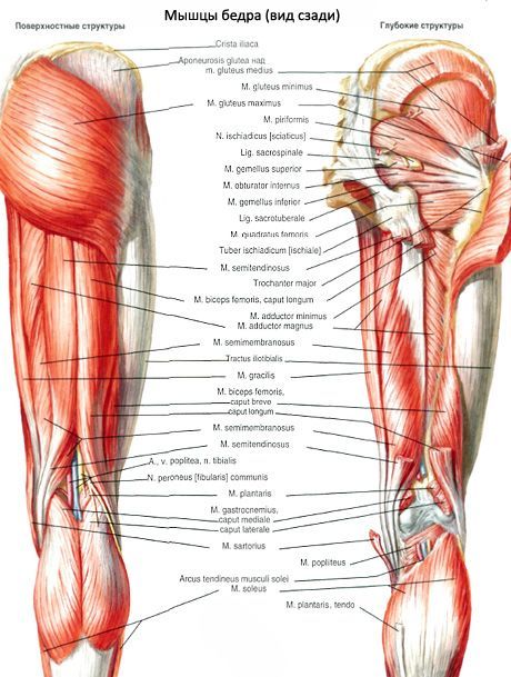
The hamstring fossa
Last reviewed: 06.07.2025

All iLive content is medically reviewed or fact checked to ensure as much factual accuracy as possible.
We have strict sourcing guidelines and only link to reputable media sites, academic research institutions and, whenever possible, medically peer reviewed studies. Note that the numbers in parentheses ([1], [2], etc.) are clickable links to these studies.
If you feel that any of our content is inaccurate, out-of-date, or otherwise questionable, please select it and press Ctrl + Enter.
The popliteal fossa (fossa poplitea) has the most complex structure, bounded from above by the tendons of the semitendinosus and semimembranosus muscles (medially) and the tendon of the biceps femoris (laterally). From below, the popliteal fossa is bounded by the heads of the gastrocnemius muscle. Under the dense popliteal fascia, which is a downward continuation of the broad fascia (of the thigh), lies the tissue in which the vascular-nerve bundle passes from top to bottom. Directly under the fascia lies the tibial nerve, deeper and inward is the popliteal vein, and the popliteal artery is located deepest and most medially. The popliteal lymph nodes and lymphatic vessels are located in the fossa. The bottom of the popliteal fossa is formed by the popliteal surface of the femur and the posterior surface of the knee joint capsule, reinforced in this place by the oblique popliteal ligament and the popliteal muscle.
The cellular space of the popliteal fossa communicates with many areas of the lower limb: the posterior muscular bed of the thigh, which in turn passes into the deep cellular space of the gluteal region. Through the adductor canal, the popliteal fossa communicates with the femoral triangle. Inferiorly, the popliteal fossa communicates with the posterior region of the leg through the cellular tissue accompanying the vascular-nerve bundle in the tibial popliteal canal, and with the lateral muscular bed of the leg through the superior musculofibular canal along the common peroneal nerve.
 [
[ What do need to examine?