Medical expert of the article

New publications
Biceps femoris muscle
Last reviewed: 07.07.2025

All iLive content is medically reviewed or fact checked to ensure as much factual accuracy as possible.
We have strict sourcing guidelines and only link to reputable media sites, academic research institutions and, whenever possible, medically peer reviewed studies. Note that the numbers in parentheses ([1], [2], etc.) are clickable links to these studies.
If you feel that any of our content is inaccurate, out-of-date, or otherwise questionable, please select it and press Ctrl + Enter.
The biceps femoris (m.biceps femoris) has two heads - long and short. The long head (caput longum) together with the semitendinosus muscle originates on the superomedial surface of the ischial tuberosity and on the sacrotuberous ligament, where the superior sac of the biceps femoris (bursa musculi bicipitis femoris superior) is located. At the level of the lower third of the thigh, the long head of the biceps femoris separates from the semitendinosus muscle and joins with the short head, passing into a flat tendon. The short head (caput breve) originates on the lateral lip of the rough line, the upper part of the lateral epicondyle and on the lateral intermuscular septum of the thigh. The common tendon of the muscle runs down the posterolateral side of the knee joint and attaches to the head of the fibula and the outer surface of the lateral condyle of the tibia. Some tendon bundles continue into the fascia of the leg. Between the tendon of the muscle and the fibular collateral ligament there is a lower subtendinous bursa of the biceps femoris (bursa subtendinea m.bicipitis femoris inferior).
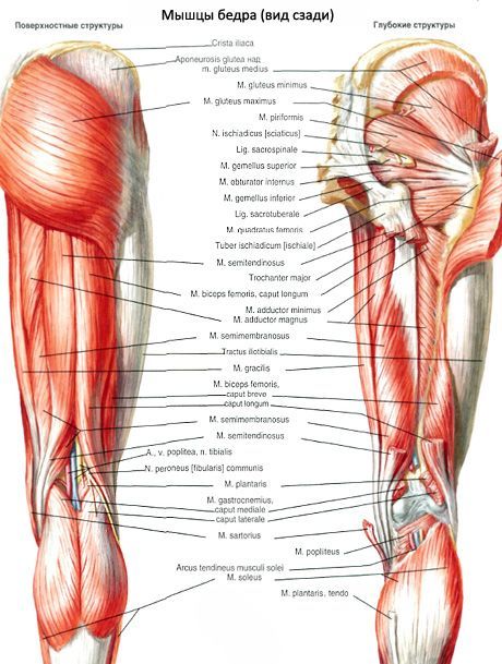
Function of the biceps femoris: together with other muscles of the posterior group, it extends the thigh; flexes the lower leg at the knee joint; when the lower leg is bent at the knee joint, it rotates it outward.
Innervation of the biceps femoris: long head - tibial nerve (SI-SII), short head - common peroneal nerve (LIV-SI).
Blood supply of the biceps femoris: medial artery, circumflex femoral artery, perforating arteries.
How to examine?