Medical expert of the article

New publications
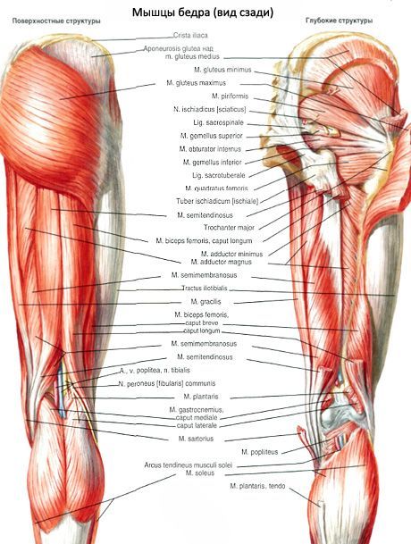
Thigh muscles
Last reviewed: 04.07.2025

All iLive content is medically reviewed or fact checked to ensure as much factual accuracy as possible.
We have strict sourcing guidelines and only link to reputable media sites, academic research institutions and, whenever possible, medically peer reviewed studies. Note that the numbers in parentheses ([1], [2], etc.) are clickable links to these studies.
If you feel that any of our content is inaccurate, out-of-date, or otherwise questionable, please select it and press Ctrl + Enter.
The thigh muscles are divided into 3 groups: anterior (hip flexors), posterior (hip extensors) and medial (hip adductors).
Having a large mass and considerable length, these muscles are capable of developing great force, acting on both the hip and knee joints. The thigh muscles perform static and dynamic functions when standing and walking. Like the pelvic muscles, the thigh muscles reach their maximum development in humans due to upright walking.
Anterior thigh muscle group
The sartorius muscle (m.sartorius) originates on the superior anterior iliac spine. The muscle crosses obliquely from top to bottom and medially the anterior surface of the thigh. It is attached, passing into a tendinous extension, to the tuberosity of the tibia and to the fascia of the leg.
The quadriceps femoris is a strong muscle, with the largest mass of all muscles. It consists of 4 muscles that form its heads: the rectus, lateral, medial and intermediate broad muscles of the thigh, which are adjacent to the femur on almost all sides. In the distal third of the thigh, all 4 heads form a common tendon that is attached to the tuberosity of the tibia, as well as to the apex and lateral edges of the patella. Distally from the apex of the patella, the middle part of the tendon continues into the patellar ligament (lig. patellae).
 [
[ Hamstrings
The posterior group of muscles includes the biceps femoris, semitendinosus, and semimembranosus. Proximally, at their origin on the ischial tuberosity, they are covered by the gluteus maximus. Below, in the posterior region of the thigh, the semitendinosus and semimembranosus muscles are located medially, adjacent to the adductor magnus. The biceps femoris occupies a lateral position and is adjacent to the vastus lateralis. Starting from the level of the border between the middle and lower thirds of the thigh, the muscles diverge to the sides, so the semitendinosus and semimembranosus muscles limit the popliteal fossa on the medial side, and the biceps femoris - on the lateral side.
The biceps femoris (m.biceps femoris) has two heads - long and short. The long head (caput longum) together with the semitendinosus muscle originates on the superomedial surface of the ischial tuberosity and on the sacrotuberous ligament, where the superior sac of the biceps femoris (bursa musculi bicipitis femoris superior) is located. At the level of the lower third of the thigh, the long head of the biceps femoris separates from the semitendinosus muscle and joins with the short head, passing into a flat tendon.
The semitendinosus muscle (m.semitendinosus) begins together with the long head of the biceps femoris on the ischial tuberosity. At the level of the middle third of the thigh, it passes into a long tendon, which runs down on the posteromedial side of the knee joint and is attached to the medial surface of the upper part of the tibia (participates in the formation of the superficial pes anserinus).
The semimembranosus muscle (m.semimembranosus) begins on the ischial tuberosity with a flat, long tendon. The tendinous plate continues downwards and, narrowing distally, passes at the level of the middle of the thigh into the muscle belly. This belly is located in front of the semitendinosus muscle and the long head of the biceps femoris. At the level of the knee joint, the muscle belly again continues into a flat tendon, which is attached by 3 bundles to the posterolateral surface of the medial condyle of the tibia. These tendinous bundles of the semimembranosus muscle form the so-called deep pes anserinus.
Medial thigh muscle group
The muscles of the medial group include the gracilis, pectineus, and adductor muscles (long, short, and large). The main function of the muscles of this group is to adduct the thigh, which is why they are called adductor muscles. They are highly developed in humans due to upright posture. These muscles originate on the outer surface of the ischium and pubis, near the obturator foramen. The origins of the muscles occupy a relatively large surface area - from the level of the pubic tubercle to the ischial tuberosity. The adductor muscles are attached in the area from the lesser trochanter to the medial epicondyle of the femur. The general direction of the muscle bundles is oblique, they pass from front to back, from top to bottom to the rough line of the femur, which serves as the attachment site for most of these muscles.
The gracilis muscle (m. gracilis) is flat, long, and located superficially along the entire length of the medial surface of the thigh. It begins with a short tendon on the lower half of the pubic symphysis and on the lower branch of the pubic bone. In the lower third of the thigh, the belly is located between the sartorius and semimembranosus muscles. The tendon of the gracilis muscle is attached to the medial surface of the upper part of the body of the tibia and participates in the formation of the superficial goose foot.
The pectineus muscle (m.pectineus) is short, flat, and originates on the crest and upper branch of the pubic bone. It is attached by a flat, thin tendon to the area located between the posterior surface of the lesser trochanter and the rough line of the thigh.
The long adductor muscle (m.adductor longus) has a triangular shape, is located medially and inferiorly to the pectineus muscle, covers the short adductor muscle and the upper bundles of the large adductor muscle in front. It begins with a thick tendon on the outer surface of the pubic bone (between the crest and the pubic symphysis). The short adductor muscle (m.adductor brevis) is thick, triangular in shape. It begins on the outer surface of the body and inferior branch of the pubic bone. It is located behind the pectineus muscle and the long adductor muscle. Directed downward and laterally, the muscle expands and is attached by short tendinous bundles to the upper part of the rough line.
Long and short adductor muscles
The large adductor muscle (m.adductor magnus) is thick, triangular in shape. It begins on the ischial tuberosity, the branch of the ischium and the inferior branch of the pubic bone. It is attached along the entire length of the medial lip of the rough line. It is located behind the short and long conducting muscles. Behind it are the semitendinosus, semimembranosus muscles and the long head of the biceps femoris. The bundles of the proximal part of the muscle are oriented almost horizontally, passing from the pubic bone to the upper part of the body of the thigh.