Medical expert of the article

New publications
Shin muscles
Last reviewed: 04.07.2025

All iLive content is medically reviewed or fact checked to ensure as much factual accuracy as possible.
We have strict sourcing guidelines and only link to reputable media sites, academic research institutions and, whenever possible, medically peer reviewed studies. Note that the numbers in parentheses ([1], [2], etc.) are clickable links to these studies.
If you feel that any of our content is inaccurate, out-of-date, or otherwise questionable, please select it and press Ctrl + Enter.
The shin muscles, like other muscles of the lower limb, are well developed, which is determined by the function they perform in connection with upright walking, statics and dynamics of the human body. Having an extensive origin on the bones, intermuscular partitions and fascia, the shin muscles act on the knee, ankle and foot joints.

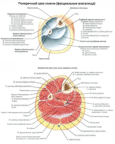
There are anterior, posterior and lateral groups of muscles of the lower leg. The anterior group includes the anterior tibialis muscle, long extensor of the fingers, long extensor of the big toe. The posterior group includes the triceps surae muscle (consisting of the gastrocnemius and soleus muscles), plantar and popliteal muscles, long flexor of the fingers, long flexor of the big toe, posterior tibialis muscle. The lateral group of the lower leg includes the short and long peroneal muscles.
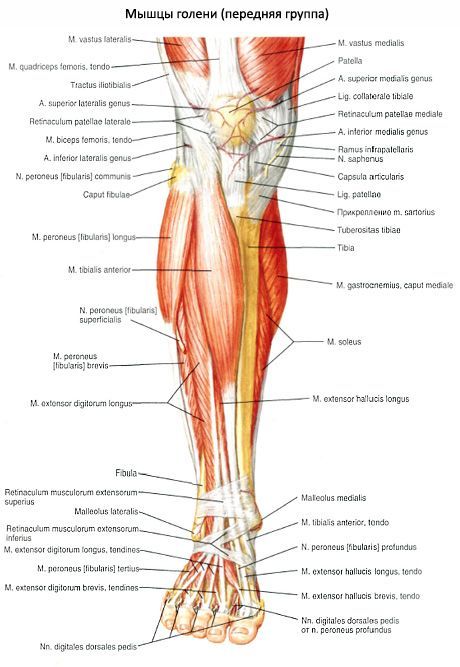
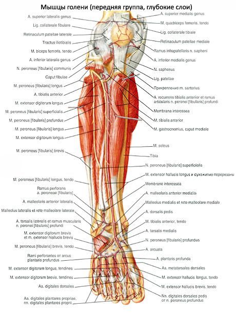
Anterior calf muscle group
The anterior tibialis muscle (m.tibialis anterior) is located on the anterior side of the leg. It originates on the lateral condyle and the upper half of the lateral surface of the body of the tibia, as well as the adjacent part of the interosseous membrane and on the fascia of the leg. At the level of the distal third of the leg, the muscle bundles pass into a long tendon, which passes under the upper and lower retainers of the extensor tendons, in front of the ankle joint. Then the tendon bends around the medial edge of the foot and attaches to the plantar surface of the medial cuneiform bone and the base of the first metatarsal bone.
Function: extends the foot at the ankle joint, simultaneously lifts the medial edge of the foot and turns it outward (supination), strengthens the longitudinal arch of the foot. When the foot is fixed, tilts the shin forward; helps keep the shin in a vertical position.
Innervation: deep peroneal nerve (LIV-SI).
Blood supply: anterior tibial artery
The long extensor of the fingers (m.extensor digitorum longus) is a pennate muscle that originates on the lateral condyle of the tibia, the anterior surface of the body of the fibula, the upper third of the interosseous membrane, the fascia and the anterior intermuscular septum of the leg. Heading to the dorsum of the foot, the muscle passes behind the upper and lower retainers of the extensor tendons. At the level of the ankle joint, the muscle divides into 4 tendons, which are enclosed in a common synovial sheath. Each tendon is attached to the dorsum of the base of the middle and distal phalanges of the II-V fingers.
A small bundle, called the third fibular muscle (m.peroneus tertius), separates from the lower part of the muscle; its tendon is attached to the base of the 5th metatarsal bone.
Function: extends the II-V toes at the metatarsophalangeal joints, as well as the foot at the ankle joint. The third fibular muscle raises the lateral edge of the foot. When the foot is strengthened, the long extensor of the fingers holds the shin in a vertical position.
Innervation: deep peroneal nerve (LIV-SI). Blood supply: anterior tibial artery.
The long extensor of the big toe (m.extensor hallucis longus) is located between the anterior tibialis muscle medially and the long extensor of the fingers laterally; it is partially covered by them in front. It originates on the middle third of the anterior surface of the fibula, the interosseous membrane of the leg. The tendon of the muscle passes down to the dorsum of the foot under the upper and lower retainers of the extensor tendons in a separate synovial sheath and is attached to the distal phalanx of the big toe. Separate bundles of the tendon can also be attached to the proximal phalanx.
Function: extends the big toe; also participates in the extension of the foot at the ankle joint.
Innervation: deep peroneal nerve (LIV-SI).
Blood supply: anterior tibial artery.
 [
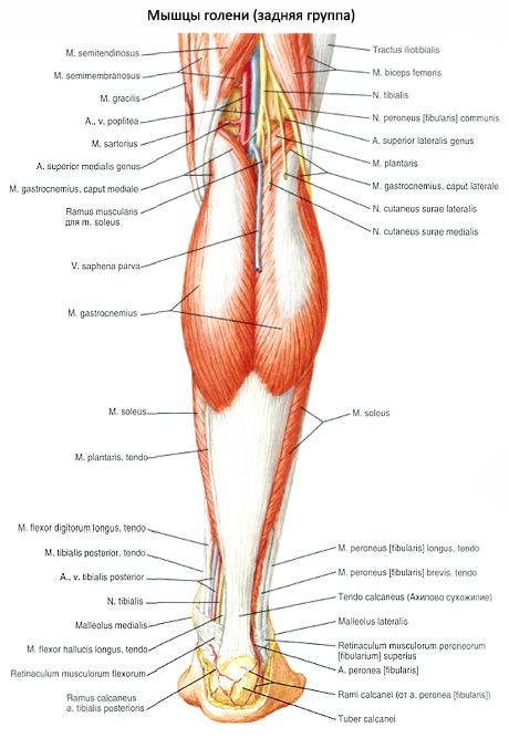
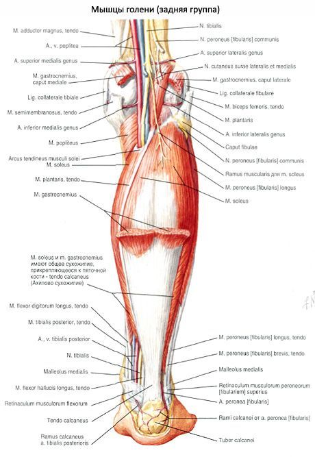
[ Posterior calf muscle group
The muscles of the posterior group form two layers - superficial and deep. The superficially lying triceps surae muscle is more strongly developed, which creates the characteristic roundness of the shin in humans. The deep layer is formed by a small popliteal muscle and 3 long muscles: the long flexor of the fingers (located most medially), the posterior tibialis muscle (occupies an intermediate position) and the long flexor of the big toe (located laterally).
Superficial layer of the posterior group of muscles of the leg
The triceps surae muscle consists of two muscles - the gastrocnemius muscle, which is located superficially, and the soleus muscle, hidden under the gastrocnemius. The gastrocnemius muscle is a two-joint muscle, it acts on two joints - the knee and ankle, while the soleus muscle is a single-joint muscle - it acts only on the ankle joint.
The gastrocnemius muscle (m.gastrocnemius) has two heads: medial and lateral, the superficial layers of which are represented by strong tendon bundles. The lateral head (caput laterale) begins on the outer surface of the lower femoral epiphysis above the lateral condyle. The medial head (caput mediate) begins on the medial condyle of the femur. Under each head of the gastrocnemius muscle is a synovial bursa. Between the lateral head and the capsule of the knee joint is the lateral subtendinous bursa of the gastrocnemius muscle (bursa subtendinea musculi gastrocnemii lateralis). Between the medial head and the joint capsule is the medial subtendinous bursa of the gastrocnemius muscle (bursa subtendinea musculi gastrocnemii medialis). Both bursae, as a rule, communicate with the cavity of the knee joint.
In the middle of the shin, both heads of the gastrocnemius muscle pass into a thick tendon, which narrows downwards and merges with the tendon of the soleus muscle, forming the calcaneal (Achilles) tendon (tendo calcaneus, s.Achilli), which is attached to the calcaneal tubercle. Between the tendon and the calcaneus there is a bursa of the calcaneal (Achilles) tendon (bursa tendinis calcanei, s.Achillis).
The soleus muscle is thick, flat, and lies under the gastrocnemius muscle. In front of it are the muscles of the deep layer. The soleus muscle has a large origin on the back surface of the tibia (on the line of the soleus muscle) and on the tendinous arch (arcus tendineus musculi solei), which is thrown between the tibia and fibula. The soleus muscle has a pennate structure, passes into a flat tendon, which participates in the formation of the Achilles tendon.
Function: The triceps flexes the leg and foot (plantar flexion); when the foot is fixed, it holds the leg on the talus, preventing it from tipping forward.
Innervation: tibial nerve (LIV-SI).
Blood supply: posterior tibial artery.
Plantar muscle
(m.plantaris) is inconstant, has a small belly and a long thin tendon. It begins on the lateral epicondyle of the femur and on the oblique popliteal ligament. The tendon of this muscle passes between the gastrocnemius and soleus muscles, adjoins the medial edge of the calcaneal tendon, together with which it is attached to the calcaneal tuberosity.
Function: stretches the capsule of the knee joint, participates in flexion of the leg and foot.
Innervation: tibial nerve (LIV-SII).
Blood supply: popliteal artery.
Deep layer of the posterior group of muscles of the leg
The deep layer is formed by 4 muscles: the popliteal muscle, the long flexor of the fingers, the long flexor of the big toe and the posterior tibialis muscle, which are separated from the soleus muscle by the deep plate of the fascia of the leg.
The popliteus muscle (m.popliteus) lies deep in the popliteal fossa. It begins with a thick tendon on the outer surface of the lateral femoral condyle (below the attachment of the fibular collateral ligament). The muscle is adjacent to the posterior surface of the joint capsule and is located below the arcuate popliteal ligament, on which its medial bundles begin. The muscle is attached to a triangular area on the posterior surface of the tibia, above the line of the soleus muscle.
Function: flexes the leg, turning it inward; stretches the capsule of the knee joint, protecting the synovial membrane from pinching.
Innervation: tibial nerve (LIV-SII).
Blood supply: popliteal artery.
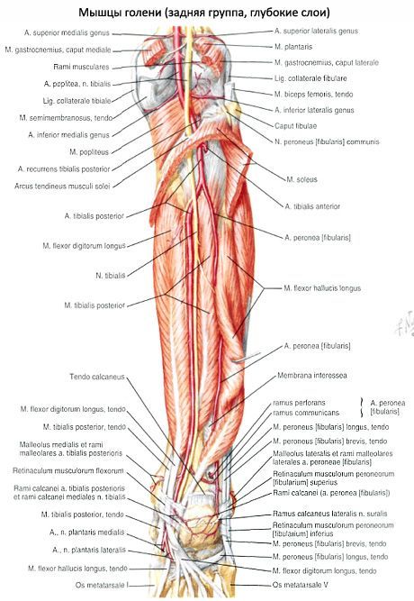
The long flexor of the fingers (m.flexor digitorum longus) has a bipinnate structure, begins with fleshy bundles on the posterior surface of the body of the tibia below the line of the soleus muscle, as well as on the fascia and posterior intermuscular septum of the leg. It is located behind and medial to the posterior tibialis muscle. The tendon of the long flexor of the fingers goes down, crosses the tendon of the posterior tibialis muscle from behind and from the lateral side. Then the tendon of the muscle passes to the sole of the foot behind the medial malleolus under the retinaculum of the flexor tendons in a separate synovial sheath (between the tendons of the posterior tibialis muscle medially and the long flexor of the big toe laterally). Then the tendon bends around the support of the talus from behind and from below. Situated above the short flexor of the fingers, it divides into 4 separate tendons that attach to the distal phalanges of the II-V fingers, having previously pierced the tendons of the short flexor of the fingers (similar to the tendons of the deep flexor of the fingers on the hand).
Function: flexes the distal phalanges of the II-V toes; flexes the foot, turning it outward.
Innervation: tibial nerve (LIV-SII).
Blood supply: posterior tibial artery.
Flexor hallucis longus
(m.flexor hallucus longus) - a bipennate muscle, originates on the lower two-thirds of the body of the fibula, the interosseous membrane, and the posterior intermuscular septum of the leg. It is located laterally and behind the posterior tibialis muscle. The tendon of the long flexor of the big toe passes under the retinaculum of the flexor tendons behind the medial malleolus and lateral to the tendon of the long flexor of the fingers in a separate synovial sheath. Then the tendon of the long flexor of the big toe lies in the groove of the same name on the posterior process of the talus, passing forward under the support of the talus. Having reached the plantar surface of the big toe, the tendon of the long flexor of the big toe is attached to its distal phalanx. On its way on the foot, this tendon crosses with the tendon of the long flexor of the fingers (lies under it). Along the entire length of the plantar surface of the first metatarsal bone, the tendon of the long flexor of the big toe lies between the medial and lateral bellies of the short flexor of the big toe.
Function: flexes the big toe, participates in flexion (supination) and adduction of the foot; strengthens the longitudinal arch of the foot.
Innervation: tibial nerve (LIV-SII).
Blood supply: posterior tibial and peroneal arteries.
The posterior tibialis muscle (m.tibialis posterior) is located deep on the back of the leg between the long flexor of the fingers (medially) and the long flexor of the big toe (laterally). It originates on the back of the body of the fibula (between the medial crest and the interosseous margin), the lower surface of the lateral condyle and on the upper two-thirds of the body of the tibia (below the line of the soleus muscle) and the interosseous membrane of the leg.
The muscle continues into a strong tendon that lies in a groove on the back of the medial malleolus in front of the tendon of the long flexor of the fingers (under the retinaculum of the flexor tendons). Passing to the plantar surface of the foot, the tendon attaches to the tuberosity of the navicular bone, to all 3 cuneiform bones, and also to the base of the IV (sometimes V) metatarsal bone.
Function: flexes the foot (plantar flexion), adducts the foot and supinates it.
Innervation: tibial nerve (LIV-SII).
Blood supply: posterior tibial artery.
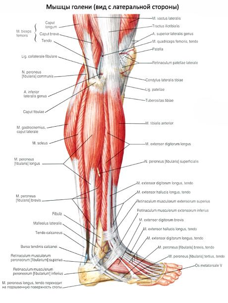
Lateral muscle group of the lower leg
The lateral group is represented by the long and short peroneal muscles, which are located on the lateral surface of the leg under the fascia between the anterior and posterior intermuscular septa.
The peroneus longus muscle (m.peroneus longus) is bipennate, lies superficially, originates on the head and upper two-thirds of the lateral surface of the fibula, on the lateral condyle of the tibia, fascia of the leg and on the intermuscular septa of the leg. At the level of the ankle joint, the tendon of the muscle, bending around the lateral malleolus from behind, passes first under the upper retainer of the tendons of the peroneal muscles in a common synovial sheath with the tendon of the peroneus brevis muscle, and then in a groove on the calcaneus (under the lower retainer of the tendons of the peroneal muscles). On the sole, the tendon of the peroneus longus muscle passes obliquely forward and medially, lies in the groove of the same name on the cuboid bone in a separate (proper) synovial sheath. The tendon attaches to the base of the first and second metatarsal bones and to the medial cuneiform bone.
At the points where the tendon changes direction (behind the lateral malleolus and on the cuboid bone), it usually thickens due to the formation of fibrocartilage or a sesamoid bone within its thickness.
Function: flexes the foot, raises its lateral edge (pronation), strengthens the transverse and longitudinal arches of the foot.
Innervation: superficial peroneal nerve (LIV-SI).
Blood supply: lateral inferior genicular artery, peroneal artery.
The peroneus brevis muscle (m.peroneus brevis) is bipennate, originating on the lower two-thirds of the lateral surface of the fibula and on the intermuscular septa of the leg. The tendon of the muscle passes onto the foot behind the lateral malleolus under the retainer of the peroneal tendons, lying in a common synovial sheath together with the tendon of the peroneus longus. At the lower edge of this retainer, the tendon of the peroneus brevis muscle turns forward and passes along the outer side of the calcaneus under the fibular block to the place of attachment at the base of the 5th metatarsal bone.
Function: raises the lateral edge of the foot; prevents the sole of the foot from turning inward; flexes the foot (plantar flexion).
Innervation: superficial peroneal nerve (LIV-SI).
Blood supply: peroneal artery.