
Medical expert of the article

New publications
Facial pigmentation: causes and methods of correction
Last updated: 11.03.2026

All iLive content is medically reviewed or fact checked to ensure as much factual accuracy as possible.
We have strict sourcing guidelines and only link to reputable media sites, academic research institutions and, whenever possible, medically peer reviewed studies. Note that the numbers in parentheses ([1], [2], etc.) are clickable links to these studies.
If you feel that any of our content is inaccurate, out-of-date, or otherwise questionable, please select it and press Ctrl + Enter.
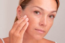
Facial pigmentation is not a separate diagnosis, but rather an external sign of increased melanin synthesis in a particular area of the skin, a change in the distribution of melanosomes, or an accumulation of existing pigment. In practice, the term "spots" most often refers to melasma, solar lentigines, and post-inflammatory hyperpigmentation, but the list is not exhaustive. Some lesions only appear as "ordinary dark spots," although in reality, they require the exclusion of melanoma and other tumors.
Melasma is considered one of the most common causes of symmetrical facial pigmentation in adults. It is more common in women and is especially common in people with darker skin phototypes. It is characterized by a chronic course, a tendency to recur, and is associated with sun exposure, hormonal fluctuations, pregnancy, oral contraception, and genetic predisposition. [1]
Solar lentigines have a different mechanism. These are localized benign spots that develop as a result of long-term ultraviolet exposure. They are more common after age 40, appearing as clearly defined, flat, light-brown or dark-brown spots and typically occurring in clusters on exposed areas, including the face. However, on the face, these spots can sometimes be difficult to distinguish from more serious pathologies without dermatoscopy. [2]
Post-inflammatory hyperpigmentation develops after any inflammation or injury to the skin. It is triggered by acne, eczema, irritation, burns, bites, aggressive peels, skin picking, and even some cosmetic procedures. This condition is especially common and lasts longer in people with darker skin, as they have higher melanocyte activity and a stronger response to inflammation. [3]
It's crucial for modern dermatology that pigmentation is enhanced not only by ultraviolet radiation but also by visible light, especially in cases of melasma and post-inflammatory hyperpigmentation. Therefore, "regular sun protection" alone is insufficient for some patients. This explains the interest in tinted sunscreens containing iron oxides, which better block visible light and reduce the risk of recurrence. [4]
Facial pigmentation is more than just a cosmetic issue. Melasma and other chronic dyschromias significantly impact quality of life, forcing people to change their habits, avoiding photographs, wearing makeup without concealer, and avoiding social situations. Therefore, a proper treatment conversation should include not only "what to apply" but also realistic expectations: lightening is usually possible, but quick and permanent results are rare, especially with melasma. [5]
Table 1. The main types of facial hyperpigmentation and their primary clinical clues.
| Option | What is typical |
|---|---|
| Melasma | Symmetrical brown spots on the forehead, cheeks, and above the upper lip |
| Solar lentigines | Single or multiple distinct flat spots on exposed areas |
| Post-inflammatory hyperpigmentation | Post-acne scars, eczema scars, trauma scars, and post-procedure scars |
| Lentigo maligna melanoma | An asymmetrical spot with uneven borders and uneven color |
| Exogenous ochronosis | Paradoxical bluish-gray darkening after long-term uncontrolled bleaching |
| Drug-induced pigmentation | Association with the use of certain drugs and more atypical coloration |
What forms are most common?
Melasma typically appears as symmetrical, light-brown to gray-brown spots and macules on the central face, cheekbones, and, less commonly, along the jawline. It is characterized by the absence of coarse scaling, ulceration, and infiltration. Clinically, it can appear quite typical, but still requires confirmation by a specialist, as similar color patterns occur in other conditions. [6]
Solar lentigines, often referred to by patients as "age spots," typically have more defined borders and appear as discrete, stable lesions against a background of chronic photodamage. Unlike melasma, they are often asymmetrical in distribution and frequently coexist with other signs of photoaging. Their primary practical significance is that they often respond well to procedural treatment, but they are also sometimes clinically confused with lentigo maligna melanoma. [7]
Post-inflammatory hyperpigmentation is characterized by the fact that it almost always leaves a mark in the area of previous inflammation. If a patient has acne along the cheekbones and jawline, the pigmentation is also distributed across these areas. The epidermal variant often appears brown, while the deeper dermal variant takes on a grayish-bluish tint and lasts significantly longer. In some people, lightening does occur spontaneously without treatment, but this process can take months or years. [8]
In everyday practice, it's equally important to be aware of the rare but crucial "masks" of hyperpigmentation. These include lentigo maligna melanoma, pigmented actinic keratosis, some forms of seborrheic keratosis, and exogenous ochronosis after prolonged, uncontrolled use of hydroquinone. If a physician or patient views any spot as "just another melasma," the risk of error increases dramatically. [9]
A separate problem is the false belief in the universality of "bleaching" products. Facial hyperpigmentation is not a single disease, but a group of conditions with varying pigment depth, different triggers, and different treatment responses. A treatment that helps with melasma may be weak against sunburn, and an aggressive procedure that quickly lightens lentigines can worsen melasma or trigger new post-inflammatory hyperpigmentation. [10]
That's why the modern approach isn't based on the principle of "the most powerful whitening," but on the principle of precise phenotyping. First, it's important to understand what exactly is present: melasma, lentigo, post-inflammatory pigmentation, or a suspicious lesion. Only then can sun protection, topical therapy, and systemic options and procedures be selected. [11]
Table 2. What most often provokes different types of facial pigmentation.
| Factor | What is it especially associated with? |
|---|---|
| Ultraviolet radiation | Melasma, solar lentigines, post-inflammatory hyperpigmentation |
| Visible light | First of all, melasma and post-inflammatory hyperpigmentation |
| Pregnancy and hormonal fluctuations | Melasma |
| Oral contraceptives and some medications | Melasma, drug-induced pigmentation |
| Acne, eczema, burn, injury | Post-inflammatory hyperpigmentation |
| Aggressive peels and incorrect procedures | Post-inflammatory hyperpigmentation |
| Chronic sun exposure | Solar lentigines |
| Uncontrolled long-term hydroquinone | Exogenous ochronosis |
When should you urgently show the stain to a doctor?
Any facial pigmentation requires prompt in-person evaluation if it is changing rapidly. The most important signs of concern are described by the rule "asymmetry, irregular border, uneven color, large diameter, and evolution." The American Academy of Dermatology specifically emphasizes that spots with irregular borders, varying shades of brown, black, white, red, or blue, as well as any lesions that change over time, are suspicious. [12]
Other warning signs include bleeding, pain, itching, crusting, hardening, ulceration, or a feeling that the spot has "changed." Benign melasma typically doesn't behave this way. Solar lentigines also typically don't suddenly bleed or ulcerate. If such symptoms appear, the doctor should consider not only pigment but also neoplasia.
A single asymmetrical spot on chronically photodamaged facial skin in an older person is particularly concerning. It is in this group that lentigo maligna melanoma can clinically mimic solar lentigo or "old harmless spot." Therefore, all slow-growing, asymmetrical dark lesions on the face should not be automatically considered benign simply because they are flat and painless. [13]
After cosmetic procedures, an expedited evaluation is necessary if, instead of gradual lightening, a sudden darkening, a gray-blue coloration, severe inflammation, persistent burning, or new uneven pigmentation appears. In such a situation, the doctor should rule out severe post-inflammatory hyperpigmentation, contact dermatitis, irritation, and, less commonly, exogenous ochronosis. The longer the incorrect approach continues, the more difficult it will be to even out skin tone. [14]
Urgency doesn't always mean life-threatening, but it does mean diagnosis is needed first, and then treatment. The main rule of thumb is simple: if a spot doesn't look typical of symmetrical melasma, changes, darkens unevenly, bleeds, hardens, or doesn't fit the overall skin pattern, it shouldn't be bleached, but rather examined by a dermatologist. [15]
Table 3. Red flags for facial pigmentation. [16]
| Sign | Why is this important? |
|---|---|
| Asymmetry | Possible melanoma or atypical pigmented lesion |
| Jagged or blurry edges | It is necessary to exclude a malignant process. |
| Uneven color | Concerns about melanoma |
| Quickly change the size or shade | Benign pigmentation doesn't usually behave like this. |
| Bleeding, crust, ulcer | An in-person dermatological evaluation is required. |
| Seal or raised component | Tumor exclusion is required |
| A single spot on an elderly person's face | Differentiation with lentigo maligna melanoma is necessary. |
Diagnostics
Diagnosis begins with a good medical history. It's important to determine when the spots appeared, whether they change between summer and winter, and whether there was pregnancy, hormonal therapy, oral contraceptive use, acne, eczema, burns, peels, lasers, or the self-administration of acids, retinoids, and bleaching agents. For post-inflammatory hyperpigmentation, prior inflammation is especially important, and for melasma, hormonal triggers and chronic sun exposure are important. [17]
The next step is a clinical examination. The dermatologist evaluates the symmetry, clarity of borders, tone, presence of scaling, vascularity, signs of photoaging, and associated rashes. Even at this stage, it is often possible to determine whether the condition is melasma, lentigo, post-inflammatory pigmentation, or a condition that cannot be reliably classified as benign. [18]
Dermoscopy is now a crucial part of assessing pigmented spots on the face. For melasma, it helps better understand the pigment distribution, and for solar lentigines and suspicious lesions, it helps determine whether there are signs of melanoma. Dermoscopy often helps avoid unnecessary biopsies in one patient and, conversely, schedule a timely biopsy for another. [19]
A Wood's lamp can be useful for understanding the depth of pigment, particularly in melasma and epidermal postinflammatory hyperpigmentation. However, it is not a perfect or definitive method. It helps to understand how superficial the pigment is and how well it might theoretically respond to topical therapy, but a diagnosis is still made based on the totality of the data. [20]
A biopsy isn't necessary for everyone. It's performed when the spot appears atypical, there's doubt between benign and malignant processes, the lesion bleeds, hardens, changes, or doesn't fit well with melasma or lentigo. This principle is especially important on the face, where a misdiagnosis is far more dangerous than a delay in aggressive cosmetic correction. [21]
A diagnosis should answer three practical questions: what type of pigmentation is it, how deep is the pigment, and are there any red flags? Only then can a specific treatment plan be discussed. Starting with strong products or procedures without this understanding is one of the most common causes of deterioration. [22]
Table 4. Which diagnostic methods are particularly useful for facial pigmentation. [23]
| Method | What is it for? |
|---|---|
| Anamnesis | Helps identify triggers, hormonal factors, inflammation, and care errors |
| Clinical examination | Allows to differentiate between typical and suspicious lesions |
| Dermatoscopy | Clarifies the structure of the spot and helps exclude melanoma |
| Wood's lamp | Helps to roughly estimate the depth of pigment |
| Serial photography | Useful for monitoring dynamics and response to treatment |
| Biopsy | Needed in case of diagnostic doubt and red flags |
Treatment
Sun protection remains the foundation of treatment for any facial hyperpigmentation. The American Academy of Dermatology recommends using sunscreen daily on exposed skin, even on cloudy days. For the face, the Academy recommends applying at least 1 teaspoon of sunscreen to the face, ears, and neck, and reapplying every 2 hours and after sweating or washing when outdoors. Without this, any depigmenting regimen is less effective and relapses more quickly. [24]
With melasma, UV protection alone is often insufficient. Research and reviews show that visible light also plays a significant role in this condition, making tinted formulas containing iron oxides advantageous. When used as a topical treatment, these products improve melasma severity better than conventional non-tinted sunscreens and reduce the risk of recurrence. [25]
The most studied topical therapy for melasma remains a combination of hydroquinone, tretinoin, and a weak corticosteroid. A fixed-dose triple combination of 4% hydroquinone, 0.05% tretinoin, and 0.01% fluocinolone acetonide has proven efficacy, and its licensed use is for short courses of up to 8 weeks, not for uncontrolled long-term use. This is crucial, as prolonged and improper use of hydroquinone increases the risk of irritation and a rare but serious complication: exogenous ochronosis. [26]
If hydroquinone isn't suitable or a gentler maintenance regimen is needed, azelaic acid, retinoids, cysteamine, and some other depigmenting agents are used. Azelaic acid is particularly useful for patients with post-inflammatory hyperpigmentation following acne, as it combines anti-inflammatory and lightening effects. Retinoids help by accelerating epidermal renewal and enhancing the effects of other treatments, but require careful administration to avoid irritation and new pigmentation.
Tranexamic acid occupies a special place. For melasma, there is evidence of the efficacy of both topical and oral formulations, particularly in patients with recurrent or resistant melasma. However, oral tranexamic acid is used off-label, requires informed consent, and is contraindicated in patients with thromboembolic risks, active thrombosis, hypersensitivity to the drug, and in women taking combined hormonal contraceptives. [27]
These treatments are not a "one-size-fits-all" quick fix. Superficial chemical peels can be an effective adjunct to melasma, but they must be gentle and used in conjunction with properly prepared skin and strict sun protection. A 2024 systematic review found that chemical peels can generally be safe and effective for melasma; however, this does not eliminate the risk of irritation and post-inflammatory hyperpigmentation, especially in darker skin types. [28]
Lasers and light-based treatments require even greater caution. For solar lentigines, they often show good results and are often superior to topical treatments. However, for melasma, lasers are not considered a first-line treatment because even with a good initial response, relapses, irritation, new post-inflammatory hyperpigmentation, and an unstable effect are possible. Therefore, melasma is first stabilized with topical therapy and photoprotection, and these procedures are considered only in carefully selected patients. [29]
Post-inflammatory hyperpigmentation is treated differently: first, the underlying inflammation is controlled. As long as the patient continues to suffer from acne, eczema, or persistent skin irritation, even effective lightening products will be incomplete. The most underestimated stage of treatment here is not addressing existing spots, but preventing the appearance of new ones. [30]
Table 5. Where which treatment is really appropriate. [31]
| Method | When it is especially appropriate | The main limitation |
|---|---|---|
| Daily photoprotection | For all forms of hyperpigmentation | Without regularity, the effect drops sharply. |
| Tinted sunscreen with iron oxides | Primarily for melasma and post-inflammatory hyperpigmentation | Application discipline is needed |
| Triple combination with hydroquinone | For moderate to severe melasma | Short course, risk of irritation |
| Azelaic acid | For post-inflammatory hyperpigmentation and sensitive skin | Works slower |
| Retinoids | As part of combination therapy | Often annoying at first |
| Tranexamic acid | For resistant melasma, as prescribed by a doctor | Oral form requires patient selection |
| Chemical peels | As an adjunct to basic therapy | Risk of inflammation and new pigment |
| Lasers and light | More often with solar lentigines, less often with melasma | Recurrence and risk of post-inflammatory hyperpigmentation |
Prevention and prognosis
Prevention of facial hyperpigmentation begins not with bleaching, but with proper skin care and protection from light. If the skin is exposed to UV and visible light daily, and the patient uses acids, retinoids, and depigmenting products irregularly, the effect will almost always be weak and unstable. This is especially true for melasma: without sun protection, it almost inevitably returns. [32]
For post-inflammatory hyperpigmentation, the main preventative principle is to treat the initial inflammation early and gently. Early control of acne, eczema, and irritating dermatoses reduces the risk of developing persistent spots. It is equally important to avoid squeezing inflammatory lesions, rubbing the skin, and using harsh home treatments. [33]
For patients with darker phototypes, prevention is especially important because their skin's pigment response is stronger and longer lasting. In this group, improper peeling, harsh scrubs, irritating skincare, or overly aggressive lasers are more likely to leave new dark marks. Therefore, the darker the initial phototype and the more active the skin's response to inflammation, the more cautious the approach should be. [34]
Uncontrolled, long-term bleaching cannot be considered preventative. Hydroquinone is effective, but it must be used according to a specific regimen, under supervision, and for a limited period. Long-term, uncontrolled use increases the risk of irritation and rare exogenous ochronosis, which is significantly more difficult to treat than the initial melasma. [35]
The prognosis depends on the type of pigmentation. Solar lentigines often respond well to treatment. Post-inflammatory hyperpigmentation often lightens slowly and requires patience, especially if the pigment is located deeper. Melasma, on the other hand, typically behaves as a chronic, recurring condition: it can be significantly lightened and stabilized, but promising complete and permanent disappearance without the risk of recurrence would be unrealistic. [36]
Table 6. What really reduces the risk of increased pigmentation. [37]
| Measure | Why does it help? |
|---|---|
| Daily photoprotection | Reduces stimulation of melanocytes |
| Tinted products with iron oxides | Provides additional protection from visible light |
| Early treatment of acne and eczema | Reduces the risk of post-inflammatory spots |
| Avoid squeezing and damaging the skin | Reduces inflammation and secondary pigmentation |
| Careful introduction of retinoids and acids | Reduces the risk of irritation |
| Avoiding long-term uncontrolled hydroquinone | Reduces the risk of ochronosis and irritation |
| A thoughtful choice of procedures with a doctor | Reduces the risk of pigment increase after peels and lasers |
FAQ
Is it possible to immediately recognize melasma just by looking at a spot?
Sometimes the clinical picture is very typical, especially if the spots are symmetrical and located on the forehead, cheeks, and above the upper lip. However, a confident diagnosis cannot be made from a photograph or a mirror, as some other conditions can look similar. This is why a dermatologist, if necessary, uses dermatoscopy, a Wood's lamp, and sometimes a biopsy. [38]
Why do spots return even after successful treatment?
Because melasma is a chronic, recurring condition that is sensitive to sun, visible light, and hormonal factors. If the spots are cleared but the triggers are not controlled and sun protection is not maintained, recurrence is very likely. This doesn't mean treatment was futile; it means melasma almost always requires a maintenance phase. [39]
Which pigmentation is easiest to treat?
Solar lentigines are often treated more predictably, especially with procedural methods. Melasma is usually more difficult because it tends to recur. Post-inflammatory hyperpigmentation can take a long time to lighten, especially if active inflammation persists or the pigment is located deeper. [40]
Does everyone need hydroquinone?
No. Hydroquinone remains an important and well-researched treatment, but it's not suitable for everyone and shouldn't be used for months without supervision. For some patients, it's better to start with gentler regimens, such as azelaic acid and retinoids. Furthermore, prolonged, improper use can cause irritation and exogenous ochronosis. [41]
Can oral tranexamic acid be used without a prescription?
No. For melasma, it is an off-label drug prescribed by a doctor after a risk assessment. Before using it, it is important to rule out active thromboembolic conditions, significant risk factors for thrombosis, and certain drug combinations, including combined hormonal contraceptives. [42]
Why is regular sunscreen sometimes insufficient?
Because not only UV radiation but also visible light plays a role in melasma and post-inflammatory hyperpigmentation. This is why tinted sunscreens with iron oxides, which provide broader protection and reduce the likelihood of recurrence, work better for some patients. [43]
When is a biopsy necessary?
A biopsy is needed not for every hyperpigmentation, but rather when there is diagnostic doubt. It is considered if the lesion is asymmetrical, has irregular borders, is uneven in color, bleeds, thickens, changes in color, or does not resemble typical melasma or lentigo. [44]
Is a laser better than creams?
It depends on the type of pigmentation. For solar lentigines, lasers often produce quick and effective results. For melasma, the situation is more complex: lasers are not considered first-line treatment because relapses and post-inflammatory hyperpigmentation remain a real problem. In such cases, photoprotection and topical therapy remain the mainstay. [45]
How long does it usually take for treatment effects to be visible?
Noticeable lightening of melasma and post-inflammatory hyperpigmentation usually develops gradually, rather than over a few days. A short course of triple combination treatments is designed for approximately 8 weeks, while more gentle regimens work more slowly. Therefore, the lack of dramatic results after 2 weeks doesn't necessarily mean the treatment is ineffective, but the plan should be reconsidered if a reasonable time has passed and there is no progress. [46]
More information of the treatment