Medical expert of the article

New publications
Celomic pericardial cyst
Last reviewed: 12.07.2025

All iLive content is medically reviewed or fact checked to ensure as much factual accuracy as possible.
We have strict sourcing guidelines and only link to reputable media sites, academic research institutions and, whenever possible, medically peer reviewed studies. Note that the numbers in parentheses ([1], [2], etc.) are clickable links to these studies.
If you feel that any of our content is inaccurate, out-of-date, or otherwise questionable, please select it and press Ctrl + Enter.
A pericardial cyst is considered a benign formation with thin walls. It can be recognized by a formation of round and irregular shape, of different diameters. In the middle of these neoplasms there is a liquid medium. It changes color and consistency under the influence of various factors. It was first described in 1852. In 1926, the first successful operation to remove a cyst from the chest cavity was performed.
Epidemiology
Of the numerous neoplasms of the middle mediastinum, cysts account for 21-22%. In 60%, the cyst is located in the cardiodiaphragmatic angular plane on the right. 30% of cysts are located on the left, and only 12% are localized at the base of the heart muscle. Women are subject to this pathology approximately three times more often than the male half of the population, which is due to the peculiarities of the anatomical structure and physiology. The peak incidence falls on the age range from 20 to 55 years.
Causes pericardial cysts
The exact cause has not been fully clarified to this day. The results of numerous scientific studies suggest that the main etiological factor is abnormal development of the pericardial sac, which occurs in the prenatal period. Transformations of the primary pericardial sheets occur predominantly. A number of genetic experiments have proven that the basis of the pericardial neoplasm develops precisely at the initial stages of fetal development. At first, these are small lacunae, which subsequently merge. There is also another theory, according to which the cyst is considered a result of abnormal development of the pleura in the prenatal period. A limited area of the pleura is separated and isolated, from which the growth subsequently forms. Gradually, it develops, fills with fluid. There is always a risk of malignant degeneration of the tumor.
The causes of tumor development in adults are traumatic injuries to the chest and heart. If there was a hematoma in a certain area, then a cyst often forms in the place of its localization. Often the cause is a tumor, then the cyst is considered one of the stages of its development. It can be provoked by inflammatory and infectious processes. Pericarditis and endocarditis lead to a cyst.
Risk factors
The high-risk group includes people with a family history of various anomalies and heart defects. The risk also increases significantly in those people who have previously been exposed to inflammatory, tumor and infectious diseases of the heart, have injuries and hematomas.
Pathogenesis
The pathogenesis is based on the disturbance of the embryonic development of the pericardium: the formation of lacunae, plates. They gradually undergo lengthening, and they are connected to each other. At first, multiple cavities are formed, gradually they merge and form a single cavity. Liquid filling occurs.
Symptoms pericardial cysts
It is mostly asymptomatic. It can often be detected only during examination of the abdominal organs. If the disease is symptomatic, patients report pain in the sternum area, accompanied by a dry cough. The intensity of pain is directly proportional to the size of the cyst. If the tumor is localized in the area of nerve passage, the pain often radiates. There is compression of the mediastinum, a person notes pain, dysphagia, shortness of breath.Cyanosis may appear. As a result, a pleuropulmonary shock condition develops.
Quite often the disease is completely asymptomatic. Therefore, if you experience any unusual sensations of discomfort, burning, pressure, you should immediately contact a specialist and undergo an examination. Also, the first symptom may be: a feeling of weakness, increased fatigue, weight loss, and other signs that could indirectly indicate a pathological process.
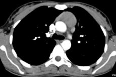
A coelomic cyst of the pericardium is characterized as a formation with thin walls and fluid inside. It forms a stalk, less often fused with the pericardial zone. It often proceeds latent, asymptomatically. Symptoms often appear if the cyst is quite large. In this case, shortness of breath, rapid heartbeat, and arrhythmia appear. A coelomic cyst can be detected using X-ray examinations, computed tomography, ultrasound echocardiography, and thoracoscopy. Treatment is only surgical.
 [ 17 ]
Complications and consequences
Diagnostics pericardial cysts
It is important to undergo preventive examinations in a timely manner. The doctor will conduct a general examination and prescribe the necessary tests.
It is difficult, but possible, to diagnose a cyst based on a visual examination. Thus, the general picture looks approximately as follows: in the place of localization and development of the tumor process, the chest protrudes. The breathing process is sharply weakened, and the affected part sharply lags behind the breathing process. During auscultation, weakened breathing is heard in the area of tumor localization. By palpation, a bony protrusion, protrusion in the chest can be detected. During percussion, a weakening of the percussion sound in the chest area can be detected, especially in the place of tumor localization.
[ 20 ], [ 21 ], [ 22 ], [ 23 ]
Tests
The main method of research is instrumental. However, analyses can be used to clarify the overall picture, identify the direction of the main processes in the body. The main analyses are clinical, biochemical blood analysis, urine and feces analysis.
The most informative is a blood test. It allows to identify the general trend of the phenomena occurring in the body. Thus, an increased ESR and an increased number of leukocytes may indicate inflammation. In the case of a tumor process, the number of lymphocytes may decrease sharply, and the ratio of the main components of the blood is also disrupted.
Instrumental diagnostics
The main method by which a cyst is detected is fluoroscopy. It is necessarily performed at various angles and taking into account various projections. A cyst is indicated by a darkening in the area of the bronchopulmonary tissue. Gradually, the darkening forms a shadow. Using this method of examination, morphological and anatomical features of the cyst structure are determined. A single-chamber tumor is smooth, a two-chamber tumor is wavy. Tomographic methods allow you to identify a cyst, distinguish it from diverticula, and detect its contour. A thin-walled chamber is visualized. If the patient turns or makes movements, a pericardial diverticulum can be seen.
Magnetic resonance imaging is also considered one of the most informative methods. It allows visualizing a tumor, distinguishing between malignant and benign tumors, as well as between inflammatory processes.
An echocardiogram in combination with an ultrasound examination is very informative.
Catheterization is an invasive procedure performed by a surgeon. Its essence lies in the invasion of the cardiac cavity in order to examine the atria and ventricles, and assess the integrity of the cardiac walls.
Thoracoscopy is an endoscopic method that allows visual detection of all neoplasms present in the heart and assessment of their parameters.
Differential diagnosis
The doctor must differentiate the cyst from tumors, diaphragmatic hernia and lipomas.
Who to contact?
Treatment pericardial cysts
If there are no complaints or concerns, treatment is not required. As soon as signs of tumor growth are detected, surgical intervention is immediately required.
Pericardial cyst removal
Today, there are two known methods: it can be removed using open abdominal surgery or thoracoscopy.
The open method is one of the most dangerous options. It is dangerous due to its complications, there are numerous contraindications. The danger is the high risk of developing massive bleeding during the operation. The risk of postoperative bleeding, infection, and other complications increases significantly. The recovery period is very long.
During thoracoscopic removal, large incisions are not made. The operation involves several main incisions, then probing is performed using a special device, which makes it possible to remove the tumor with minimal damage, targeted. It is completely isolated, so there is no re-growth. The risk of bleeding, infection is practically absent. The person recovers much faster. In general, the algorithm of the operation can be presented as follows: first, an incision is made and the cyst is carefully examined, then the doctor begins to enucleate it. This ends with complete removal. If the cyst is too large and filled with air, it is removed from the liquid media.
Treatment of pericardial cysts with folk remedies
There are no folk or medicinal remedies. The only way to treat the disease is surgical intervention, in which the tumor is removed.
Prevention
Prevention of cysts comes down to timely diagnostics. Regular preventive examinations are necessary. If diseases of the circulatory system or concomitant diseases are detected, they must be completely cured, which will reduce the risks. It is necessary to avoid injuries and damage.
[ 26 ]
Forecast
If you start treating it in time, the prognosis is favorable - the pericardial cyst can be completely removed and will stop bothering the person. The recovery period, as a rule, is quite easy. If treatment is not carried out, the further course of events can be extremely unfavorable, even fatal.