Medical expert of the article

New publications
Cardiac catheterization
Last reviewed: 06.07.2025

All iLive content is medically reviewed or fact checked to ensure as much factual accuracy as possible.
We have strict sourcing guidelines and only link to reputable media sites, academic research institutions and, whenever possible, medically peer reviewed studies. Note that the numbers in parentheses ([1], [2], etc.) are clickable links to these studies.
If you feel that any of our content is inaccurate, out-of-date, or otherwise questionable, please select it and press Ctrl + Enter.
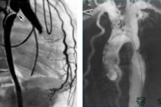
Catheterization of the cardiac cavities is performed using puncture and percutaneous insertion of a catheter into a vessel - a peripheral vein (ulnar, subclavian, jugular, femoral) for the right chambers of the heart or an artery (brachial, femoral, axillary, radial) for the left chambers of the heart.
 [
[ Methodology for performing cardiac catheterization
In case of severe narrowing of the aortic valve or its artificial prosthesis, when it is impossible to retrogradely pass a catheter into the left ventricle, a transseptal puncture of the interatrial septum from the right atrium to the left and then into the left ventricle is used. The most frequently used approach to the vessel is according to the Seldinger method (1953). After local anesthesia of the skin and subcutaneous tissue with 0.5-1% novocaine solution or 2% lidocaine solution and a small notch on the skin, a vein or artery is punctured with a needle; when blood appears from the proximal tip of the needle (pavilion) (it is necessary to try to puncture only the anterior wall of the vessel), a guidewire is inserted through the needle, the needle is removed and a catheter is passed into the vessel along the guidewire, which, naturally, should be longer than the catheter. The catheter is advanced to the required location under X-ray control. In case of using floating catheters of the Swan-Gans type with a balloon at the end, the location of the catheter tip is determined by the pressure curve. It is preferable to install a thin-walled introducer with a hemostatic valve and a side branch for washing into the vessel, and through it it is easy to insert the catheter and replace it with another one if necessary. The catheter and introducer are washed with heparinized isotonic sodium chloride solution to prevent thrombus formation. Using different types of catheters, it is possible to reach different parts of the heart and vessels, measure pressure in them, take blood samples for oximetry and other tests, introduce RVC to determine anatomical parameters, constrictions, blood discharge, etc.
If there is no fluoroscopic (X-ray) control over the location of the catheter, catheters with an inflatable floating balloon at the end are used, which can move with the blood flow into the right atrium, right ventricle, pulmonary artery and record the pressure in them. The pulmonary artery wedge pressure allows indirectly judging the state of the left ventricle function, its end-diastolic pressure (EDP), since the left ventricular EDP is the average pressure in the left atrium or the pressure in the pulmonary capillaries. This is important for monitoring therapy in cases of hypotension, heart failure, for example, in acute myocardial infarction. If the catheter has additional devices, it is possible to measure cardiac output using dye dilution or thermodilution, record an intracavitary electrogram, and perform endocardial stimulation. The intracavitary pressure curves are recorded using a Statham liquid pressure sensor and an ECG on a jet recorder or computer with possible printing on paper; their changes can be used to judge a particular cardiac pathology.
Measurement of cardiac output
It should be noted that there are no absolutely accurate methods for measuring cardiac output. During cardiac catheterization, three methods for determining cardiac output are most often used: the Fick method, the thermodilution method, and the angiographic method.
Fick's method
It was proposed by Adolph Fick in 1870. The method is based on the assumption that at rest, the oxygen supply to the lungs is equal to the amount of oxygen utilized by the tissues, and the amount of blood ejected by the LV is equal to the volume of blood flowing through the lungs. Mixed venous blood must be taken, since the oxygen concentration in the blood of the vena cava and coronary sinus differs significantly. Blood is taken from the RV or pulmonary artery, which is preferable. The arteriovenous oxygen difference can be determined from the oxygen concentration in arterial (Ca) and venous (Cv) blood. By calculating the oxygen content absorbed during 1 min, the volume of blood flowing through the lungs during the same period of time can be calculated, i.e., the cardiac output (CO):
MO = Q / Ca - St (l/min),
Where Q is the oxygen absorption by the body (ml/min).
Knowing the cardiac index, you can calculate the cardiac index (CI). To do this, divide the cardiac index by the patient's gel surface area, which is calculated based on his height and body weight. The cardiac index in an adult is normally 5-6 l/min, and the CI is 2.8-3.5 l/min/ m2.
Thermodilution method
This method uses a cooled isotonic sodium chloride solution (5-10 ml), which is introduced through a multi-lumen catheter into the right atrium, the tip of the catheter with the thermistor is in the pulmonary artery. Calibration of the curves is carried out by briefly switching on a constant resistance, which gives deviations of the recording device corresponding to a certain change in temperature for a given thermistor. Most thermodilution devices are equipped with analog computing devices. Modern equipment allows for up to 3 measurements of blood MO within 1 min and multiple repetitions of the studies. Cardiac output, or MO, is determined by the following formula: MO = V (T1 - T2) x 60 x 1.08 / S (l/min),
Where V is the volume of the introduced indicator; T1 is the blood temperature; T2 is the indicator temperature; S is the area under the dilution curve; 1.08 is the coefficient depending on the specific density and heat capacity of the blood and isotonic sodium chloride solution.
The advantages of thermodilution, as well as the need for catheterization of only the venous bed, make this method currently the most acceptable for determining cardiac output in clinical practice.
Some technical aspects of the catheterization laboratory
The staff of the catheterization angiography laboratory includes the head, physicians, operating nurses and X-ray technicians (X-ray technicians) if cine and large-format X-ray filming is used. In laboratories using only video films and computer image recording, X-ray technicians are not needed. All laboratory staff must be proficient in cardiopulmonary resuscitation techniques, for which the X-ray operating room must have the appropriate medications, a defibrillator, a device for electrical stimulation of the heart with a set of electrode catheters, a central oxygen supply and (preferably) an apparatus for artificial ventilation of the lungs.
Complex and risky diagnostic procedures and PCI (angioplasty, stenting, atherectomy, etc.) should preferably be performed in clinics with a cardiac surgery team. According to the recommendations of The American College of Cardiology/American Heart Association, angioplasty and examination of patients with a high risk of complications, AMI can be performed by experienced, qualified specialists without the presence of cardiac surgery support in the hospital if the patient cannot be transported to a more suitable location without additional risk. In Europe and some other countries (including Russia), endovascular interventions are increasingly performed without the presence of cardiac surgeons, since the need for emergency cardiac surgery is currently extremely low. An agreement with a nearby cardiovascular surgery clinic is sufficient for emergency transfer of the patient there in case of peri- and postprocedural complications.
To maintain the fitness, qualification and skill of the operators, the laboratory must perform at least 300 procedures per year, and each physician must perform at least 150 diagnostic procedures per year. For catheterization and angiography, a high-resolution X-ray angiography unit, a system for monitoring ECG and intravascular pressure, archiving and processing angiographic images, sterile instruments and various types of catheters are required (different types of catheters for coronary angiography are described below). The angiography unit must be equipped with an attachment for cineangiographic or digital computer image acquisition and archiving, have the ability to obtain images online, i.e. immediately with quantitative computer analysis of angiograms.
Changes in intracavitary pressure curves
Intracavitary pressure curves may change in various pathological conditions. These changes serve for diagnostics when examining patients with various cardiac pathologies.
To understand the causes of pressure changes in the cavities of the heart, it is necessary to have an idea of the temporal relationships between the mechanical and electrical processes occurring during the cardiac cycle. The amplitude of the a-wave in the right atrium is higher than the amplitude of the y-wave. An excess of the y-wave over the a-wave in the pressure curve from the right atrium indicates a violation of the filling of the atrium during ventricular systole, which occurs with tricuspid valve insufficiency or a defect
In tricuspid stenosis, the right atrial pressure curve resembles that in the left atrium in mitral stenosis or constrictive pericarditis, with a decline and plateau in mid- and late diastole, typical of elevated pressures during early systole. The mean left atrial pressure corresponds fairly closely to the pulmonary artery wedge pressure and the pulmonary trunk diastolic pressure. In mitral insufficiency without stenosis, there is a rapid decline in pressure at the onset of systole (a decrease in the y-wave), followed by a gradual increase in late diastole (diastasis). This reflects the achievement of equilibrium between atrial and ventricular pressures during the late phase of ventricular filling. In contrast, in patients with mitral stenosis, the y-wave decreases slowly, while the pressure in the left atrium continues to decrease throughout diastole, and there are no signs of diastasis of the pulse pressure in the left atrium, since the atrioventricular pressure gradient is preserved. If mitral stenosis is accompanied by a normal sinus rhythm, the α-wave in the left atrium is preserved and contraction of the atria causes the creation of a large pressure gradient. In patients with isolated mitral regurgitation, the v-wave is clearly expressed and has a steep descending knee of the y-line.
On the left ventricular pressure curve, the EDP point immediately precedes the onset of its isometric contraction and is located immediately after the a-wave before the c-wave of left atrial pressure. Left ventricular EDP may increase in the following cases: heart failure, if the ventricle experiences a large load caused by excessive blood flow, for example, in aortic or mitral insufficiency; left ventricular hypertrophy, accompanied by a decrease in its distensibility, elasticity and compliance; restrictive cardiomyopathy; constrictive pericarditis; cardiac tamponade caused by pericardial effusion.
In aortic valve stenosis, which is accompanied by obstructed blood outflow from the left ventricle and an increase in pressure in it compared to the systolic pressure in the aorta, i.e. the appearance of a pressure gradient, the left ventricular pressure curve resembles the pressure curve during isometric contraction. Its outlines are more symmetrical, and the maximum pressure develops later than in healthy individuals. A similar picture is observed when recording pressure in the right ventricle in patients with pulmonary artery stenosis. Blood pressure curves may also differ in patients with different types of aortic stenosis. Thus, in valvular stenosis, a slow and delayed increase in the arterial pulse wave is observed, and in hypertrophic cardiomyopathy, the initial sharp increase in pressure is replaced by a rapid decrease and then a secondary positive wave reflecting obstruction during systole.
Derived indices of intraventricular pressure
The rate of change/increase of the intraventricular pressure curve during the isovolumic contraction phase is called the first derivative - dр/dt. Previously, it was used to assess the contractility of the ventricular myocardium. The value of dр/dt and the second derivative - dр/dt/р - are calculated from the intraventricular pressure curve using electronic and computer technology. The maximum values of these indicators represent the indices of the ventricular contraction rate and help to assess the contractility and inotropic status of the heart. Unfortunately, the wide range of these indicators in different categories of patients does not allow us to develop any average standards, but they are quite applicable in one patient with initial data and against the background of the use of drugs that improve the contractile function of the heart muscle.
Currently, having in our arsenal of patient examination methods such as echocardiography in its various modifications, computer (CT), electron beam and magnetic resonance imaging (MRI), these indicators for diagnosing cardiac pathologies are not as important as before.
Complications of cardiac catheterization
Cardiac catheterization is relatively safe, however, like any invasive technique, it has a certain percentage of complications associated with both the intervention itself and the general condition of the patient. The use of more advanced and thin atraumatic catheters, low-osmolar and/or non-ionic RVS, modern angiographic units with real-time computer image processing for invasive interventions has significantly reduced the incidence of possible complications. Thus, mortality during cardiac catheterization in large angiographic laboratories does not exceed 0.1%. S. Pepine et al. report an overall mortality rate of up to 0.14%, with 1.75% for patients under 1 year of age, 0.25% for people over 60, 0.03% for single-vessel coronary artery disease, 0.16% for three-vessel disease, and 0.86% for left coronary artery disease. In case of heart failure, mortality also increases depending on the NUHA class: in I-II FC - 0.02%, III and IV FC - 0.12 and 0.67%, respectively. In some patients, the risk of serious complications is increased. These are patients with unstable and progressive angina, recent (less than 7 days) myocardial infarction, signs of pulmonary edema due to myocardial ischemia, with circulatory failure of III-IV FC, severe right ventricular failure, valvular heart defects (severe aortic stenosis and aortic regurgitation with a pulse pressure of more than 80 mm Hg), congenital heart defects with pulmonary hypertension and right ventricular failure.
In a multivariate analysis of 58,332 patients, predictors of serious complications were severe congestive heart failure, hypertension, CHD, aortic and mitral valve disease, renal failure, unstable angina and acute myocardial infarction in the first 24 hours, and cardiomyopathy. In 80-year-old patients, mortality during invasive diagnostic procedures also increased to 0.8%, and the incidence of vascular complications at the puncture site reached 5%.